Download
1 / 15
180 likes | 819 Vues
Protein modification. 16-1. Protein modifications different types, including co-translational and post-translational inteins. It is estimated that the human proteome consists of ~300,000 different proteins, or about 10X more than the number of genes (!)

E N D
Protein modification 16-1 • Protein modifications • different types, including co-translational and post-translational • inteins It is estimated that the human proteome consists of ~300,000 different proteins, or about 10X more than the number of genes (!) - protein modifications - differential splicing
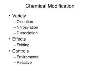
Protein modifications: formation or breakage of covalent bonds 16-2 • disulfide bond formation - covered • addition of a small moiety • phosphorylation, glycosylation, methylation, hydroxylation, etc. - today • truncation • self-cleavage - viral capsid protease (SCP) - covered - intramolecular chaperones - covered • cleavage of signal sequences - signal peptidases • protein splicing: inteins – covered in this lecture • intermolecular cleavage (e.g., by proteasome) - presentation • addition of ubiquitin or ubiquitin-like protein - presentation usually specified by amino acids at certain positions, or various degenerate or specific motifs found in proteins
N- and C-terminal modifications: acylation, methylation, amidation 16-3 • frequently, the N-terminal methionine is not present in mature proteins • Acylation includes acetylation, formylation, pyroglutamylation, myristoylation • ~80% eukaryotic cytosolic proteins are acetylated at their N-termini • makes N-terminal (Edman) sequencing difficult without special treatment - requires enzymatic removal or treatment with chemicals that may cleave labile peptide bonds • formyl group occurs mostly as modification of the initiator methionine in bacteria • pyroglutamate represents a cyclic amide generated from an N-terminal glutamic acid or glutamine residue - can be generated by spontaneous cyclization but could also be an artifact of protein isolation under slightly acidic conditions • myristoylation is a co-translational lipid modification that is common to many signalling proteins; occurs only on N-terminal glycine residues
formyl transferase (FMT) peptide deformylase (PDF) methionine aminopeptidase (MAP)
N- and C-terminal modifications: methylation and amidation 16-4 • Methylation of N-terminal amino groups is rare; different methylases do modify specific proteins, including ribosomal proteins • methylation of ribosomes affects their function • Amidation of peptides (e.g., hormones) sometimes occurs at the C-terminus
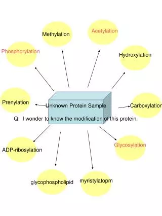
Modification of individual side chains:phosphorylation and glycosylation 16-5 • Phosphorylation • phosphorylation can affect the activity and structure of proteins • perhaps as many as 1 in 8 proteins are phosphorylated • too many examples to list: e.g. HSF activity is modulated by phosphorylation; cell-signalling molecules are best characterized • Glycosylation • glycosylation takes place in the ER, golgi by a variety of enzymes • glycosylated proteins often found on the surface of cells or are secreted • folding/assembly of glycosylated proteins requires ER molecular chaperones • addition of GlcNAc (beta-O-linked N-acetylglucosamine) residues occurs in the cytoplasm and nucleus - modifications are carried out by O-linked GlcNac transferases (OGTs) - proteins modified by O-GlcNAc include: cytoskeletal proteins, hormone receptors, kinases & other signalling molecules, nuclear pore proteins, oncogenes, transcription factors, tumor suppresors, transcriptional & translational machinery, viral proteins
Modification of individual side chains:various others 16-6 • Prenylation, fatty acid acylation • proteins without major hydrophobic (transmembrane) domains can be directed to membranes by prenylation of their C-terminal cysteine residue • Hydroxylation and oxidation, carboxylation • a variety of derivatives are known; e.g., hydroxyamino acids (hydroxyproline) are very common in collagen • Selenocysteine/selenomethionine modification • essentially all selenium in cells occurs as selenocysteine • selenomethionine is a useful too for protein crystallography: can grow cells in the presence of the modified amino acid and produce protein containing se-Met; can deduce ‘phase’ of protein this way
Identification of modifications Fig. 1. O-GlcNAc is an abundant modification of nucleocytoplasmic proteins. Nucleo-cytoplasmic proteins from HeLa cells were immunopurified with an O-GlcNAc-specific antibody and stringently washed, and the O-GlcNAc-containing proteins were specifically eluted with free GlcNAc. The resulting proteins were separated on two-dimensional gels and visualized by silver staining. pI, isoelectric point; MW, molecular weight. From Wells et al. (2001) Science291, 2376-8. use 2D gel electrophoresis to detect modified proteins in whole-cell (or partly purified) lysates 16-7 - mass spectrometry is the most common today: - can identify modifying group with great precision, especially in combination with proteolytic digestion of proteins and HPLC analysis - incorporation of radioactive groups by addition to growing cells e.g., 75Se-labeling and chromatographic isolation of proteins Note that most of the modifiers can be purchased in radiolabeled form - antibody cross-reactivity: e.g., antibody against phosphotyrosine small-molecule modifications can affect not only the activity, but also the structure of proteins, much as ligands such as ATP can affect the activity and structure of proteins
Presentation: proteasome-mediated co-translational protein biogenesis 16-8 Lin et al. (1998) Cotranslational biogenesis of NF-kappaB p50 by the 26S proteasome. Cell92, 819-828.
Protein splicing: inteins 16-9 • Inteins represent the protein equivalent to the genomic DNA intron: elements that are spliced out of the final (mature) product • unlike introns, inteins are self-splicing through the use of an endonuclease • used commercially in biochemical applications (explanation on board) - inteins are 134-608 aa - the mini-inteins do not have the endonuclease domain but have other characteristics of inteins Legend: Schematic illustration of protein splicing (upper part) and intein structure (lower part). The two terminal regions of the intein sequence form the splicing domain of a typical bifunctional intein. Six conserved sequence motifs (A to G) are shown. The intein sequence begins with the first amino acid of motif A and ends with the second last amino acid of motif G. Motifs C and E are the dodecapeptide motifs of endonuclease. A star (*) stands for hydrophobic amino acids (V, L, I, M). A dot (.) stands for a nonconserved position. adapted from Liu, X.-Q. (2000) Annu. Rev. Gen.34, 61.
Protein splicing: inteins 16-10 Legend: Scenarios of intein evolution. A, loss of the endonuclease domain. B, breaking the intein sequence. C, loss of the splicing function. D, replacing the C-extein with a cholesterol molecule (green dot). E, loss of C-terminal cleavage and splicing. F, loss of N-terminal cleavage and splicing. G, placing intein fragments on two ends of a protein. H, breaking intein into 3 fragments. I, separating a middle fragment of the intein from the rest. J, presence of two different split inteins. Scenarios A to D are based on examples observed in nature. Scenarios E to G are based on engineered artificial inteins. Scenarios H to J are purely hypothetical.
Intein protein splicing: mechanism 16-11 • N-extein represents the N-terminal polypeptide segment that is retained • C-extein represents the C-terminal segment that is retained • the Intein is what is spliced out (much as a genomic DNA intron) • Cys1, Asn154 and Ser155 represent conserved residues involved in the splicing reaction
Hedgehog (Hh)processing cholesterol (A) catalytic residues of the Hh protein and (B) the crystal structure of the spliced protein - modification of SH by cholesterol is required for its activation - process similar to intein splicing Hall et al. (1997) Crystal Structure of a Hedgehog Autoprocessing Domain: Homology between Hedgehog and Self-Splicing Proteins. Cell 91, 85-97. 16-12 Secreted signaling proteins encoded by the hedgehog gene family induce specific patterns of differentiation in a variety of tissues and structures during vertebrate and invertebrate development. All known signaling activities of Hh proteins reside in Hh-N; Hh-C is responsible for both the peptide bond cleavage and cholesterol transfer components of the autoprocessing reaction.
Legend to right-hand Figure, part (A) 16-13 Intramolecular Autoprocessing Reactions of Hh and Self-Splicing Proteins (A) Schematic drawing of a two-step mechanism for Hh autoprocessing (Porter et al., 1996b ). Aided by deprotonation by either solvent or a base (B1), the thiol group of Cys-258 initiates a nucleophilic attack on the carbonyl carbon of the preceding residue, Gly-257. This attack results in replacement of the peptide bond between Gly-257 and Cys-258 by a thioester linkage (step 1). The emerging -amino group of Cys-258 likely becomes protonated, and an acid (A) is shown donating a proton. The thioester is subject to a second nucleophilic attack from the 3 hydroxyl group of a cholesterol molecule, shown here facilitated by a second base (B2), resulting in a cholesterol-modified amino-terminal domain and a free carboxy-terminal domain. In vitro cleavage reactions may also be stimulated by addition of small nucleophiles including DTT, glutathione, and hydroxylamine.