Download
1 / 42
430 likes | 967 Vues
The Heart and blood vessels and circulation Chapter 12 and 13. I. The heart and the circulatory system. A. Blood flows through a network of blood vessels that extend between the heart and the peripheral tissues.

E N D
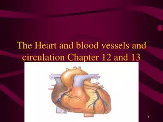
The Heart and blood vessels and circulation Chapter 12 and 13
I. The heart and the circulatory system A. Blood flows through a network of blood vessels that extend between the heart and the peripheral tissues.
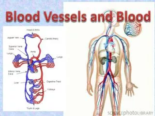
1. Blood vessels are subdivided into a pulmonary circuit, which carries blood to and from exchange surfaces of the lungs and a systemic circuit, which transports blood to and from the rest of the body.
2. Arteries, or efferent vessels carry blood AWAY from the heart. 3. Veins or afferent vessels carry blood TO the heart. 4. Capillaries are small, thin walled vessels between the smallest arteries and veins.
5. The thin walls of the capillaries permit the exchange of nutrients, dissolved gases, and waste products between the blood and surrounding tissues. 6. The heart is a small organ, the size of a clenched fist. 7. The heart consists of four chambers.
8. The right atrium receives blood from the systemic circuit and the right ventricle discharges blood into the pulmonary lungs. 9. The left atrium collects blood from the pulmonary circuit, and the left ventricle ejects it into the systemic circuit.
10. When the heart beats, the atria contract first then the ventricles. The two ventricles contract at the same time and eject volumes of blood.
B. The Anatomy and Organization of the Heart 1. The heart lies directly behind the sternum. 2. The heart is surrounded by the pericardial cavity. 3. The lining of the pericardial cavity is a serious membrane called the pericardium.
4. The heart is a hollow, muscular organ that contracts at regular intervals, forcing blood through the circulatory system.
5. The walls of the heart are made up of three layers of tissue. a. The outer and inner layers are epithelial tissues, which cover and protect other tissues. b. The middle layer is cardiac muscle tissue.
6. Cardiac muscle tissue has a rich supply of blood. 7. The cells make up cardiac muscle tissue are loaded with mitochondria. (why?)
8. The right side of the heart pumps blood from the body into the lungs where oxygen-poor (deoxygenated) blood gives up carbon dioxide and picks up the oxygen. 9. The left side of the heart pumps oxygen-rich (oxygenated) blood from the lungs to the rest of the body.
10. The heart is enclosed in protective sac of tissue called the pericardium. 11. Dividing the right side from the left side is a septum or wall. The septum prevents the mixing of oxygen-poor and oxygen –rich blood.
12. On each side of the septum are two chambers. The upper chambers, which are called atria, receive blood coming into the heart. 13. The lower chambers, which are called ventricles, pump blood out of the heart.
C. Internal anatomy and organization 1. Each atrium communicates with the ventricle on the same side through an atrioventricular (AV) valve. 2. The valve ensures a one-way flow of blood from the atria into the ventricles.
3. The right atrium receives blood from two large veins, superior vena cave and the inferior vena cava. 4. The superior vena cava delivers blood from the head, neck upper limbs, and chest. 5. The inferior vena cava carries blood from the rest of the trunk, the viscera and the lower limps.
6. The cardiac veins of the heart return venous blood to the coronary sinus, which opens into the right atrium. 7. Blood travels from the right atrium into the right ventricle through an opening bounded by three flaps. 8. These flaps or cusps are part of the AV value, they are also known as the tricuspid valve.
9. Blood leaving the right ventricle flows into the pulmonary trunk. 10. Blood flows into the left and right pulmonary arteries. These vessels branch out. 11. The left atrium has a valve the left atrioventricular valve or bicuspid valve. Also called the mitral valve.
D. Differences between right and left ventricles. 1. The function of an atrium is to collect blood returning to the heart and deliver that blood to the attached ventricle.
II. The heart beat A. The alternating contraction and relaxation of the heart chambers is called the cardiac cycle. 1. The heart does not contract in a single motion. 2. Once the ventricles contract, the blood must be directed out through the arteries and not back up into the atria.
3. Once blood has entered the arteries, it must be prevented from flowing back as the heart relaxes. 4. Pressure in one direction opens the valves, while reverse pressure forces the valves tightly closed.
5. Two seminar valves allow blood to enter the pulmonary artery and the aorta when the ventricles contract but prevent it from returning as the ventricles relax. 6. The contraction of the heart is initiated and coordinated by a pacemaker. A cluster of specialized muscle cells that produce spontaneous electrical signals at a regular rate.
7. The heart’s primary pacemaker is the sinoatrial (SA) node, located in the wall of the right atrium. 8. When a pacemaker fails to coordinate muscle contractions, a condition known as fibrillation occurs. 9. Fibrillation of the ventricles is soon fatal because blood is not pumped out of the heart to the brain and other organs but is merely sloshed around.
B. Blood vessels 1. Three types of blood vessels that are involved in the circulatory system are arteries, capillaries and veins. 2. Arteries carry blood away from the heart, In general, the walls of arteries are thicker than veins.
3. This helps arteries withstand the high pressure of blood. 4. Except for pulmonary arteries, all arteries carry oxygen rich blood. 5. The artery that carries oxygen-rich blood from the left ventricle to all parts of the body is the aorta.
6. The aorta is the largest artery in the body. As the aorta leaves the heart it continues to branch into smaller arteries and finally to arterioles. 7. Arterioles branch into networks of very small blood vessels called capillaries.
8. The flow of blood moves from capillaries into veins. Veins form a system that collects blood from every part of the body and carries it back to the heart. The smallest veins are called venues.
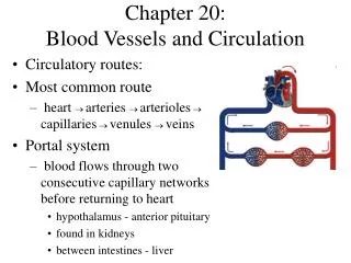
C. Pathways of circulation 1. There are two pumps involved in the circulatory system. One of the two pumps is called the pulmonary circulation. 2. This consists of the heart and lungs. The circulation begins at the right ventricle and ends at the left ventricle.
3. Oxygen depleted blood is pumped out of the right ventricle into the lungs through the pulmonary arteries. These are the only arteries that carry deoxygenated blood. 4. Blood returns to the heart through the pulmonary veins. The only veins that carry oxygen rich blood.
5. The other pump, consisting of the left atrium and ventricle, powers systemic circulation. Starts at the left ventricle and ends at the right atrium. 6. Newly oxygenated blood from the lungs enters the left atrium through pulmonary veins and is passed to the left ventricle.
III. Blood pressure A. A measure of the force that blood exerts against a vessel wall is called blood pressure. 1. The body regulates blood pressure in two ways.
3. When blood pressure is too low, the autonomic nervous system constricts the walls of the arteries. This reduces their diameter and helps to raise blood pressure. 4. When blood pressure is high, the kidneys remove more water from the blood, lowering the total amount of fluid in the circulatory system. The loss of fluid from the blood lowers the blood pressure.
4. Problems can arise when blood pressure is either too low or too high. 5. Low blood pressure slows down the rate at which blood flows through the body. The part of the body that area far away from the heart, hands and feet do not receive enough blood. This is called hypotension.
6. High blood pressure or hypertension occurs when blood pressure is high, the heart works much harder to pump blood, causing the heart muscle to weaken. 7. People with high blood pressure are also more likely to develop problems in the arteries outside the heart.
8. It is not known what causes high blood pressure but it is known certain things raise ones chance of high blood pressure. Things such as obesity, and family back ground. 9. Medicine can help to regulate blood pressure along with exercise, diet and not smoking.
10. Blood pressure can be measured by a sphygmomanometer. 11. Blood pressure is recorded as two numbers separated by a slash. 12. The number on the top is the systolic pressure. This is when the heart is working.
13. The bottom number is the diastolic pressure. This is when the heart is NOT working, or when it is done. 14. An average blood pressure for a 20 year old is 120/70.