Download
1 / 28
340 likes | 1.07k Vues
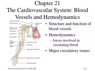
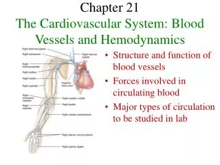
Chapter 19 – Blood Vessels and Circulation . Types of blood vessels. Arteries Carry blood away from the heart Branch into smaller vessels called arterioles Capillaries Smallest vessels Form beds in tissues/organs Where diffusion occurs Veins Carry blood back to heart

E N D
Types of blood vessels • Arteries • Carry blood away from the heart • Branch into smaller vessels called arterioles • Capillaries • Smallest vessels • Form beds in tissues/organs • Where diffusion occurs • Veins • Carry blood back to heart • Venules – small veins from capillary beds • Converge to form larger veins
Vessel walls • With the exception of capillaries, vessels have 3 layers/tunics surrounding the central lumen • Tunica intima/interna • Endothelium – simple squamous epithelium continuous with the endocardium of the heart • Tends to be convoluted in large arteries • Tunica media • Smooth muscle and elastin fibers • Can alter lumen size • Vasodilation – muscle relaxes/lumen enlarges • Vasoconstriction – muscle contracts/lumen decreases • Thicker in arteries • Tunica externa or adventitia • Loose connective tissue and collagen • Large vessels have small vessels within externa called vasa vasorum
Arterial system • Elastic/conducting arteries • Largest in diameter • Aorta and major branches • Large amount of elastic fibers • Expand and recoil • Muscular arteries • Relative to size, largest tunic media • Ability to vasocontrict • Arterioles • Few elastic fibers • Diameter controls amount of blood delivered to capillary beds
Capillaries • Consist only of tunica intima and basement membrane • Continuous – endothelium is complete • Prevents loss of blood cells and proteins • Fenestrated – contains pores • Allows for movement of small proteins • Choroid plexi, GI tract for absorption, kidneys, endocrine system • Forms beds – network of capillaries
Capillary microcirculation • Blood from arteriole to venule • Terminal arteriole → metarteriole → true capillaries → thoroughfare channel → post capillary venule • Precapillary sphincter • Band of smooth muscle at metarteriole/capillary junction • When contracted, blood bypasses capillary bed • Blood goes directly from metarteriole to thoroughfare channel
Venous system • Venules • Very porous, like capillaries • Veins • Walls are thinner and lumens are larger as compared to same size artery • Relatively little smooth muscle • Large veins tend to collapse in histological preparation • Externa is the largest layer
Venous system cont • Since veins are exposed to relatively low pressure: • Veins contain valves • Prevents backflow of blood due to gravity • Skeletal muscles “milk” veins • Contraction of muscles close to veins help move blood through the vessel • Abdominal pressure differences due to breathing • Abnormalities • Varicose veins • Poor circulation causes valves to become leaky • Veins stretch out and become floppy • Usually superficial veins that have little support from underlying tissues • Hemorrhoids – varicose veins of anal veins
Vascular anastomoses • Where multiple vessels unit • Arterial anastomoses • Arteries that supply same area merge • Provides alternate blood supply • Venous anatomoses • More common than aterial
Disorders • Atherosclerosis • Lipid deposits on arterial walls • Can cause restriction or blockage of blood flow • Arteriosclerosis • Decreased elasticity “hardening of arteries” • Affects proper blood flow
Circulation physiology • Blood flow • Volume of blood that passes through a specific point of a vessel in a specific time • ml/min • Blood pressure • Force per unit exerted by blood on vessel wall • mm Hg • Usually refers to arterial pressure • Pressure gradient – pressure difference is required for blood to flow
Circulation physiology cont • Resistance • Opposition to flow; measures friction the blood encounters • Influenced by: • Blood viscosity • Blood vessel length • Longer the vessel = greater resistance • Blood vessel diameter • Blood next to the wall flows more slowly • F = ∆P/R • F = flow; ∆P = pressure difference; R = resistance • As pressure increases, flow increases • As pressure decreases, flow decreases
Systemic blood pressure • Vessel pressure is highest in the aorta and decreases to zero in right atrium • Blood flows from region of high pressure to low • Blood pressure is measured in arteries – exposed to highest pressure • Systole – ventricular contraction • ~120mm Hg • Diastole – ventricular relaxation • ~80mm Hg
Maintaining blood pressure • Hormonal control (discussed during endocrine system) • Baroreceptors – detect pressure changes • Located in aortic arch, carotid arteries, and large head/neck arteries • When relaxed, the medulla oblongata sends signals to vessels for vasoconstriction, which increases pressure • When stretched, medulla doesn’t send signal – causes vasodilation, which decreases pressure
Maintaining blood pressure cont • Chemoreceptors – detect changes in respiratory gases • Increases in carbon dioxide is detected by aortic arch and carotid sinuses • Vasocontriction causes an increase of blood pressure, which increases blood flow • Gets to respiratory system to unload carbon dioxide more quickly • Decrease of carbon dioxide causes vasodilation – causes slower flow
Pulse • Surges of pressure in an artery • Pulse rate should equal the heart rate (beats per minute) • Apical pulse – actual heartbeat count • BIG difference = pulse deficit • Felt with fingers (do not use thumb)
Blood pressure • Pressure from blood against the vessel walls (arteries) • Reported as diastolic pressure over systolic pressure • Measured with a sphygmomanometer • Inflation blocks bloodflow through brachial artery • Pressure of cuff gradually reduced to allow partial flow • Sounds of Korotkoff– tapping sound of blood flowing back into artery • First appears at systole • Blood spurting into artery • Sound disappears when artery is no longer compressed • Diastole
Blood pressure alteration • Hypertension • >140 systole; >90 diastole • To pump against increased pressure, heart works harder • Myocardium increases (especially LV), heart ultimately weakens • Hypotension • <100 systole
Circulatory shock • Blood vessels inadequately filled; abnormal blood flow • Hypovolemic • Significant blood loss • Vascular • Poor circulation due to extreme vasodilation • Anaphylactic – allergies; septic – infection • Cardiogenic • Heart not pumping (myocardial infarction)