Download
1 / 39
390 likes | 412 Vues
Dive into the world of vision, exploring the physical stimulus of light, eye structures, contrast sensitivity, visual acuity, and the fundamental limits of human vision. Learn about the properties and measures of light, eye anatomy, receptor types, and more. Discover the fascinating mechanisms underlying our ability to perceive the world through our eyes.

E N D
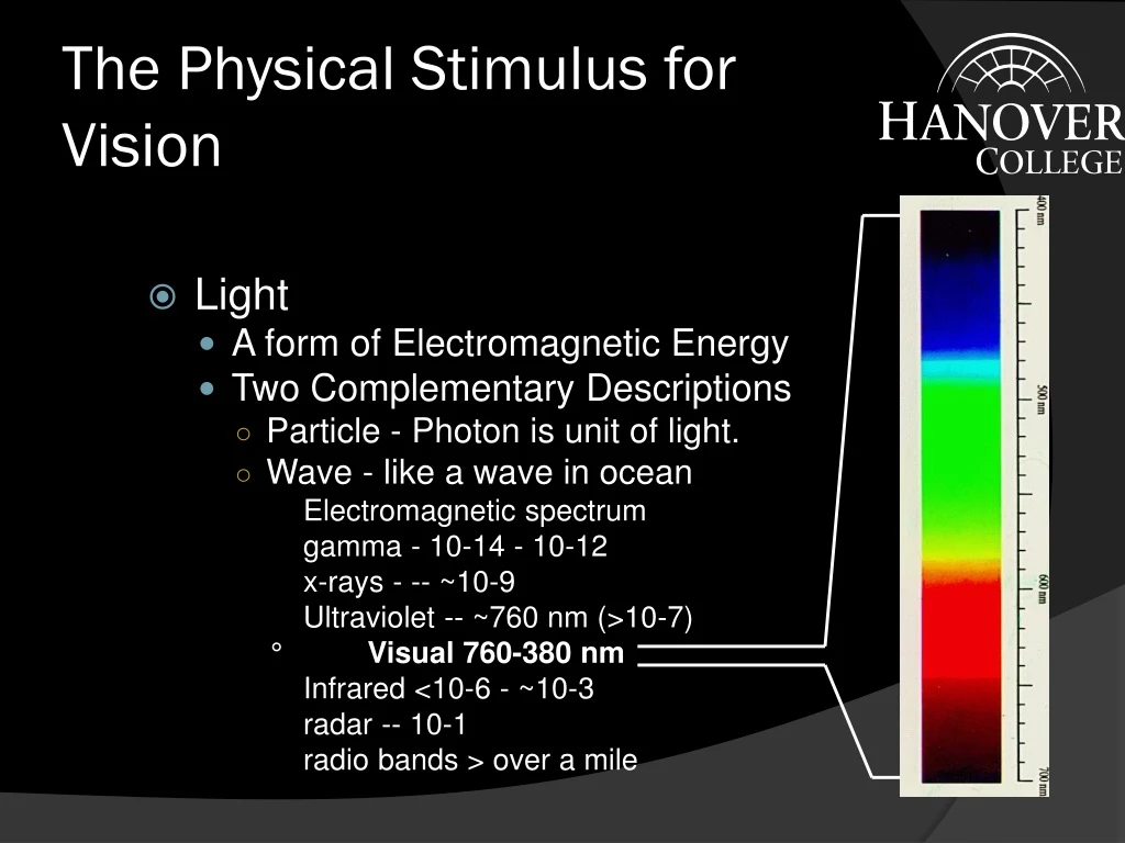
The Physical Stimulus for Vision • Light • A form of Electromagnetic Energy • Two Complementary Descriptions • Particle - Photon is unit of light. • Wave - like a wave in ocean Electromagnetic spectrum gamma - 10-14 - 10-12 x-rays - -- ~10-9 Ultraviolet -- ~760 nm (>10-7) ° Visual 760-380 nm Infrared <10-6 - ~10-3 radar -- 10-1 radio bands > over a mile
Measures of Light • Wavelength relates to color, e.g., the spectrum • Usually measured in billionths of a meter - nanometers or nm
Measures of Light - 2 • Amplitude = Intensity relates to brightness • Measures use only that light that is effective in stimulating the human eye. • Important types of measures of Amplitude 1.Illuminance - light falling on a surface 2. Luminance - light coming off a surface 3. Reflectance = luminance/illuminance 4. Contrast Ratio = luminance of brightest area/luminanceof darkest area
Structures of the Eye 1. Sclera 2. Cornea 3. Aqueous Humor 4. Iris 5. Pupil 6. Lens 7. Ciliary muscle 8. Retina 9. fovea (pit) 10. blind spot/optic disc 11. Pigment Epithelium
The Retina • Two Types of Receptors • Rods • ~120 million/eye • night vision • no color • not in fovea • most about 20deg in periphery • Cones • ~7 million/eye • day vision • three types so color vision • most in fovea from Lewis, Zeevi, & Werblin (1969)
The Retina - 2 Cones Rods Data from Osterberg (1935).
Mach Bands Click to add successively lighter bars. Watch the edge to the right side of the last bar.
Craik-Cornsweet: Described The figure above is an exaggerated map indicating the light levels across the image on the previous slide. Note how the center and edges have identical luminance. That can be seen by sitting far enough away from the screen
Minimal Contours Described There are two circles below. Both circles have the same luminance (intensity level) at the center. Click on your mouse and This one changes abruptly watch as the edges are blurred to the level at the center. and the circle disappears.
Accommodation • DEFINITION: The adjusting of the lens thickness to focus at different distances. • Necessary because can only see clearly one distance at a time • Goes very rapidly • Closes can focus in Near Point • Farthest can focus is Far Point • Loose ability to focus as age - moves towards far point • In dark accommodation moves to ~1 meter from face • As fatigue, accommodation moves to this dark focus.
Acuity • DEFINITION: ability to resolve or see fine details. • Visual Angle: DEFINITION: Angle formed by object on retina. • Types of Acuity: what is meant by acuity depends upon the stimulus used to measure it. • Detection: black bar on white field • Resolution: a grating • Recognition: e.g. Snellen, where you read letters.
Acuity - 2 • Measures of Acuity • 20/20: can see at 20’ what a normal person can see at 20’. • This is normal, not perfect, vision. 20/200: can see at 20’ what a normal person can see at 200’. • Visual angle of the critical feature in a test, e.g. the width of the bars in a grating. • A typical population average is 1 arcmin (1/60 degree). • Acuity and Retinal Location: • Best at fovea. Falls off rapidly in periphery. Is tied to density of cones.
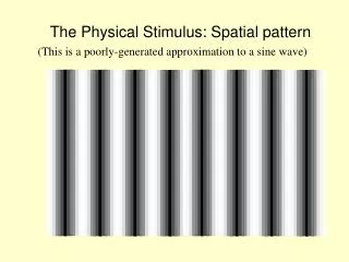
Contrast Sensitivity • DEFINITION: the minimum contrast ratio that can be detected. • Indicates the smallest difference between shades of gray that can be detected. • Depends on Spatial Frequency • DEFINITION: how many pairs of white and black bars fit into 1 deg. of visual angle. • Low spatial frequency few bars/deg. • High spatial frequency many bars/deg.
The Contrast Sensitivity Function • Our sensitivity to contrast depends on this spatial frequency. • Peak sensitivity is 4-6 cycles/degree. • The highest spatial frequency we can see at any contrast is limited by our acuity.
Contrast Sensitivity and Performance • Increasing contrast above threshold will allow for faster identification, up to a point • Beyond a certain contrast ratio - about 3 to 1 or 4 to 1 - increasing contrast ratio has no effect (Krantz, Silverstein, & Yeh, 1992)
Fundamental Limits of Vision • Operating Range of Vision - 1:1014 if: 1 cent (threshold) =100 Most incomes are between $10,000 and $100,000 = 106 to 107 GNP of U.S.A. for 1 year = ~1015
Fundamental Limits of Vision - 2 • Duplex Theory of Vision • Two eyes in one. One for day. One for night.
Dark/Light Adaptation • Dark Adaptation • The term applied to the increased sensitivity that occurs when we enter a region of lowered sensitivity. • Two phases: • early rapid phase - lasts ~7 minutes and due to cones. • later slower phase - complete in about 30 to 40 minutes due to rods. • Light Adaptation • Takes 2-3 minutes
Eye Movements • We move our eyes because of the limited field with good acuity. • There are 6 eye muscles • 4 rectus • 2 oblique • Types of Eye Movements • Version: Both eyes move together • Vergence: Eye move in opposite directions from Kaiser, 1997, http://www.yorku.ca/research/vision/ eye/thejoy.htm
Eye Movements - 2 • Version • Saccades, most common (link to ESP) • places object on fovea. • can be > 400 deg/sec. • Takes ~ 200 msec to begin • Smooth Pursuit • track moving objects • relatively slow ~30 deg/sec. • Vergence: • Convergence (together) and divergence (apart) • Allows us to look at closer and farther objects. • relatively slow and also takes about 200 msec to begin
Color Vision I: Color Matching • Dimensions of Color • Hue: refers to the color name we apply. • Saturation: purity of color, to desaturate add white • Brightness: • Trichromatic Theory of Vision • We have three classes of cones • L or red: peak at 564 nm • M or green: peak at 534 nm • S or blue: peak at 420 nm
Color Vision I: Color Matching - 2 • Color Matching in the Trichromatic Theory • Two patches of light will appear the same if the activity across the three cones is the same, regardless of wavelengths making up the two patches. • In general, can match any one color with three other colors • This is where we get three primaries. • Neutral colors - whites and grays • a balance of activity across the three receptors.
Color Vision I: Color Matching - 3 • Formalization of the Trichromatic Theory • By CIE originally in 1931 • Based on 300 observers to develop standard observer. • A set of Equations that allow predictions of matching. • Used in photo printing, TV and film. • Updates in 1960, 1976
Color Vision II: Color Deficiencies • Most can be understood using Trichromatic Theory • Dichromatism: Missing one of the three cones (link) Dichromats tend to see through camouflage better than thrichromats • Other Types • Monochromatism: One cone or only rods • Anomalous Trichromats: Three cones but one is different.
Color Vision III: Color Appearance • Color Opponent Theory • Four Primaries: red-green, blue-yellow • Arranged in opposition pairs • Red vs. Green • Blue vs. Yellow • Add on to other get neutral color • Never see a mixture of opposition pairs. • Evidence: • complimentary colors, color aftereffect, simultaneous contrast, color naming - try it with just red, green blue and yellow • Cells in visual system respond this way.
Color Vision IV: Resolution modified from Kaiser, 1997, http://www.yorku.ca/research/vision/ eye/thejoy.htm
Depth Perception • If retinas are flat (2 dimensional) how do we see depth (the 3rd dimension)? • We use cues: sources of information about depth. • Monocular or one eye cues
Depth Perception - 2 • Binocular or two eye cues • Vergence(Only cue to give absolute depth information): • muscular feedback from effort to converge or diverge gives information about depth. • works only for relatively near objects: <20’ • Stereopsis (link) • Binocular Disparity: measure of difference of position of an object on the two retinas • DEFINITION: ability to use binocular disparity to see depth. • Basis of 3-D movies • Accuracy of Depth Judgments: • In general, more cues more accurate.
Depth Perception - 3 • Size Constancy • DEFINITION: seeing objects as a relatively constant size despite change in retinal image size. Sretinal image a 1/distance to object (a mean proportional to) Sperceived = Constant • Can be quite useful in object recognition • A Variation is Emmert’s Law for after effects Sretinal image = Constant Sperceiveda distance • Also applies to depth generated by stereopsis
Depth Perception - 4 • To experience Emmert’s Law fixate the center of the dot below for about 45 seconds. Then quickly view the next slide and note the size. Then look at surfaces of different distances, also noting the size.