THE AMAZING EYE
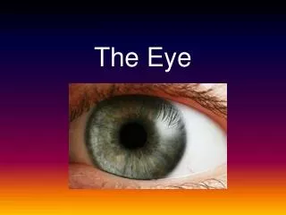
THE AMAZING EYE. THE EYE. The sense of sight, or vision , is the most complex sensory system of the body. Accessory structures to the eye include the eyelids , lacrimal apparatus , and extrinsic muscles , and the eyeball itself are located within the orbit of the skull.


THE AMAZING EYE
E N D
Presentation Transcript
THE EYE • The sense of sight, or vision, is the most complex sensory system of the body. • Accessory structures to the eye include the eyelids, lacrimal apparatus, and extrinsic muscles, and the eyeballitself are located within the orbit of the skull.
ACCESSORY STRUCTURES • The eyelids protect the anterior surfaces of the eyes. • Each eyelid consists offourlayers: • a thin outer layer • a skeletal muscle layer • a connective tissue layer • and an inner mucous membrane called the conjunctiva.
The conjunctiva folds back to cover much of the anterior surface of the eye as well. • It secretes mucus to help moisten and lubricate the eyeball.
LACRIMAL APPARATUS • The lacrimal apparatus associated with each eye consists of the lacrimal gland,lacrimal sac, and nasolacrimal ducts. • The lacrimal gland secretes tears, which are a dilute salt solution that is released continuously.
Tears also contain an antibacterial enzyme, which fights against infection as the eye is moistened and lubricated. • As the tears flush across the surface of the eye, they are collected by lacrimal canals that channel into the lacrimal sac located within the lacrimal bone. • They then pass into the nasal cavity by way of the nasolacrimal duct.
The extrinsic muscles of the eye are skeletal muscles that move the eyeball. • They originate from the walls of the orbit and insert upon the tough outer surface of the eyeball. • There are six extrinsic muscles,
EXTRINSIC MUSCLES • Lateral rectus: moves eye laterally • Medial rectus: moves eye medially • Superior rectus: elevates or rolls eye upward • Inferior rectus: depresses or rolls eye downward • Inferior oblique: elevates and turns eye laterally
STRUCTURE OF EYE • The eye is a spherical structure about 2.5 cm in diameter. Its wall consists of three distinct layers, or tunics. • The outer fibroustunic, the middlevascular tunic, and theinner nervous tunic. • Inside the eye are structures that divide the eye into fluid-filled compartments.
FIBROUS TUNIC • thick outermost layer of the eyeball. It contains two regions: • posterior sclera • anterior cornea
SCLERA • forms most of the fibrous tunic. • It is composed of white fibrousconnective tissue and is often called the “white of the eye.” • It forms a thick, tough protective layer that provides shape to the eyeball and protects its inner parts.
It contains an abundant supply of blood vessels, some of which may come into view when the eyes are irritated. • The posterior surface of the sclera is penetrated by the optic nerve.
CORNEA • The cornea is the anterior, transparent part of the fibrous tunic that bulges outward slightly. • It is the “window” of the eye, as light must pass through it before entering the internal structures and cavities. • Its transparency is due to its lack of blood vessels and its regular arrangement of protein fibers.
VASCULAR TUNIC • so called because of its abundance of blood vessels, which supply nourishment to numerous structures of the eye. • Its most prominent components include, theciliary body, and theiris. • associated with the vascular tunic is thelens.
CHOROID • a thin, dark-brown membrane that lines most of the internal surface of the sclera. • the brown pigment absorbs light • blood vessels within the choroid serve to nourish the retina, which lies internally.
Anterior to the choroid, the vascular tunic is modified to form two structures composed of smooth muscle: • the ciliary body • the iris
CILIARY BODY • The ciliary body continues from the choroid to become the thickest part of the vascular tunic. • It consists of smooth muscle fibers, which connect to the lens by way of suspensory ligaments. • Attached to the inner margin of the ciliary body is the doughnut-shaped iris
IRIS • beautifully colored part of the eye that can be seen from the exterior. • suspended between the cornea and the lens • contraction of its muscle fibers changes the diameter of the opening in its center to regulate the amount of light enteringthe inner eye cavity, called the pupil.
PUPIL • appears as a black spot in the center of your eye.
LENS • located immediately behind the pupil and iris. • in young, healthy eyes it is perfectly transparent • it is composed of strands of transparent cytoplasm that originate from epithelial cells. • the lens is held in place by suspensory ligaments.
When tension is relaxed, the elastic capsule expands and the lens becomes more convex. • This occurs when the lens focuses on a close object. • When tension is increased, the capsule flattens and the lens becomes less convex. Occurs when focused on distant object.
The alteration of lens shape for near and far vision is called accommodation. • The lens provides a physical separation between the two main compartments of the eye.
MAIN COMPARTMENTS • The anterior comp. is divided by theirisinto two chambers: • the anterior chamber, between the cornea and the iris • the posterior chamber, between the iris and the lens.
Within these two chambers a clear, watery fluid called the aqueous humor circulates and is continuously recycled through the bloodstream. • The posterior compart. behind the lens is known as the posterior cavity. • It contains a thickened, gel-like fluid that helps support the structure of the eyeball, known as the vitreous humor.
Unlike the aqueous humor, the vitreous humor is not continuously recycled.
NERVOUS TUNIC • The nervous tunic consists of the retina, which is a thin, fragile layer of neurons that forms the inner lining of the eyeball’s posterior wall. • The retina contains its own supply of blood vessels. • Function of the retina is the detection of light and subsequent transport of nerve impulses to the optic nerve, which penetrates it.
RETINA • The retina consists of three distinct layers of neurons. • The layer nearest the choroid contains the neurons that are specialized to respond to light, the photoreceptor cells.
PHOTORECEPTOR CELLS • Two types: • rod cells, each of which contains an elongated, cylindrical dendrite • cone cells, whose dendrites are tapered to a point like cones. • Rod cells are not color-sensitive, but are sensitive to very small levels of light (night vision)
Cone cells, are color-sensitive and require more light but provide a sharper image. • Both cells respond to light by initiating a signal, which is transmitted to the cells forming the middle layer of the retina, the bipolar neurons.
OPTIC DISC • point near the center of the retina, where axons from the ganglion cells converge to form the optic nerve. • called the “blind spot”
PATHWAY OF LIGHT THROUGH THE EYE • The bending of light rays is called refraction. • accommodation: the ability to change its shape in order to move the visual focus, • myopia: the amount of refraction is too great (or the eyeball is anatomically too long) and an image is focused in front of the retina, distant objects are blurred.
the individual is said to be nearsighted because close objects can still be focused normally.
HYPEROPIA • the image focuses behind the retina • the individual is farsighted, because distant objects can still be focused on properly. • Hyperopia results from a weak or “lazy” lens or an eyeball that is too short. • Unequal curvatures of the cornea or lens lead to astigmatism, which causes blurred vision for near and far objects.
image is inverted • and reversed from left to right
PHYSIOLOGY OF VISION • Rods are extremely sensitive to very small amounts of light. Therefore, are useful in night or dim vision. • They are not color-sensitive. • Cone cells are sensitive to color • In rods, the pigment is called rhodopsin, or visual purple. • Rhodopsin is composed of a protein and a substance called retinene.
Retinene: synthesized from vitamin A. • A deficiency of vitamin A in the diet leads to a condition called nightblindness, due to a decline in sensitivity of rod cells and their subsequent inability to respond to dim light.
OPTIC CHIASMA • The nerves from portions of each eyeball cross just anterior to the pituitary gland at the base of the brain, forming the X-shaped optic chiasma • Both optic tracts extend to the thalamus. • From the thalamus, the impulses enter visual pathways that lead to the occipital lobes of the cerebral cortex.
THE END ! • These centers, called the visual cortex, are where interpretation of visual signals takes place.