The Eye
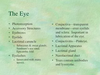
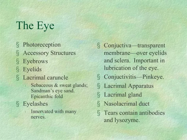
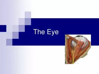
The Eye. Versatile Adaptable Strong Durable Hollow More sophisticated than the best camera!. Muscles of the Eye oculomotor muscles. Levator palpebrae superioris Eyelid muscle. Orbital fat Cushions and insulates the eye. Palpebrea Eyelid Lashes


The Eye
E N D
Presentation Transcript
Versatile Adaptable Strong Durable Hollow More sophisticated than the best camera!
Muscles of the Eye oculomotor muscles
Levatorpalpebraesuperioris Eyelid muscle Orbital fat Cushions and insulates the eye
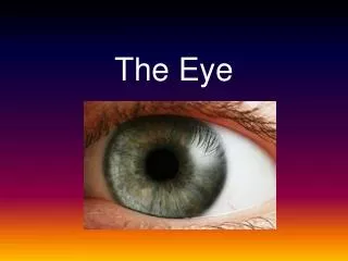
Palpebrea Eyelid Lashes Large sebaceous glands (called tarsal glands) along the inner side, like other hair follicles secrete a lipid-rich substance that keep the eyelids from sticking together. Canthus Medial and later corners of the eye
Conjunctiva Mucus covering of the eye. Begins at the eye lids and cover the outer surface of the eye. Nerve endings and is sensitive – pink eye - conjunctivitis Lacrimal apparatus canal, sac , glands, pores Produces, distributes and removes tears. Tears contains an enzyme that kills bacteria. Along with the tarsal glands, tears make an oily covering to reduce friction and evaporation.
Eye Cavities and Layers
Posterior Eye Cavity Vitreous Chamber Contains vitreous body, gelatin-like fluid Anterior Eye Cavity The front of the eye subdivided into Anterior chamber And Posterior chamber
Layers are called Tunics Page 304 Outer – fibrous tunic Sclera and cornea Middle - vascular tunic Iris, cilliary body and choroid Inner – neural tunic retina, fovea centralis
Anterior Chamber of the Eye Two chambers Anterior Cornea to the iris Posterior Iris, ciliary body and lens