The Eye
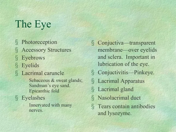
Photoreception Accessory Structures Eyebrows Eyelids Lacrimal caruncle Sebaceous & sweat glands; Sandman’s eye sand. Epicanthic fold Eyelashes Innervated with many nerves. Conjuctiva—transparent membrane—over eyelids and sclera. Important in lubrication of the eye.

The Eye
E N D
Presentation Transcript
Photoreception Accessory Structures Eyebrows Eyelids Lacrimal caruncle Sebaceous & sweat glands; Sandman’s eye sand. Epicanthic fold Eyelashes Innervated with many nerves. Conjuctiva—transparent membrane—over eyelids and sclera. Important in lubrication of the eye. Conjuctivitis—Pinkeye. Lacrimal Apparatus Lacrimal gland Nasolacrimal duct Tears contain antibodies and lysozyme. The Eye
The Human Eye • Cornea—contains many nerve endings.—transplantation and repair. • pupil • iris • Lens--convex • suspensory ligaments • ciliary bodies • Extrinsic muscles • retina • cones and rods • optic nerve • Choroid coat—Why is it dark colored?
Electromagnetic Radiation Visible light Reflection Refraction Real image Convex lens Upside down and reversed form left to right. Light and Image Formation
Focal point Focal length Retina Photoreceptors Optic disc Blind spot Optic nerve Rods cones Macula lutea Fovea centralis Forms a pathway for light to get to the photoreceptors. Most cones are in the fovea. This is where important area for hard focusing. Small portion of our field of view. Image Formation
Human Eye Defects • Myopia • Causes • Eye too long or lens too convex • Effects • Correction—concave lens. • Hyperopia • Causes • Eye too short or lens too flat • Effects • Correction—convex lens. • Presbyopia
Diplopia—movements of the extrinsic muscles of the 2 eyes are not coordinated. Leads to double vision. Strabismus—”cross-eyed” Affected eye rotates either medially or laterally. Treatments and effects. Retinal Detachment Glaucoma—excessive aqueous humor—Intraocular pressure. Cataract—clouding of the lens. May be caused by sunlight, diabetes, smoking, vitamin deficiencies,, etc… Astigmatism Color blindness Night blindness Eye Disorders
Visual Pathways to the Brain • From the retina, visual impulses travel to: • Optic nerve • Optic chiasma • Optic tracts • Thalamus • Optic radiation tract • Primary visual cortex in the optic lobe.
What is sound? Wavelength Frequency Pitch Intensity Amplitude Loudness Decibels Above 90 db is dangerous Hearing range—20 to 20000 Hz Hearing
Mechanoreceptors Fluids Outer, middle, and inner ear Outer ear Pinna or auricle Are bigger ears better? External auditory canal Ceruminous glands Tympanic membrane Middle Ear Pharyngotympanic tube Eustachian tube How does this structure function? Ossicles Malleus, incus, stapes Malleus is attached to the eardrum. Stapes is attached to the oval window of the inner ear. Sound amplification Hearing
Transmit vibrations of the eardrum to the middle ear. Inner ear Bony labyrinth Semicircular canals Vestibule Cochlea Membranous labyrinth Inside the bony labyrinth Filled with fluid Vestibule—oval window is attached to this structure. Contains equilibrium receptors. Semicircular canals—poster and lateral to the vestibule. Ampulla—equilibrium receptors. Hearing
Cochlea—anterior to the vestibule. Cochlear duct Basilar membrane Organ of Corti Hair cells Cochlear nerve Thalamus Auditory cortex in temporal lobes Disorders of the Ear Otitis Media Deafness Conduction Deafness Otosclerosis—stapes becomes fused to oval window. Sensorineural deafness—damage to hair cells or neural pathways. Cochlear implants Tinnitus Hearing
Importance of head movements. Inputs from the inner ear, eyes, and stretch receptors Vestibular apparatus Vestibule—static equilibrium Semicircular canals—dynamic equilibrium Maculae—respond to straight line changes in speed and direction, not to rotation Hairs cells Vestibular nerve Respond only to changes in velocity or acceleration. Equilibrium
Equilibrium • Crista ampullaris • Semicircular canals • Respond to rotation • Changes in rotation • Hair cells • To balance centers in the brain stem or the cerebellum • Motion sickness • Sensory input mismatch
Chemoreceptors Aqueous chemicals Taste buds Most on the tongue Papillae—projections of the surface of the tongue. Gustatory and taste cells—taste receptors Taste pore Gustatory hairs Replaced every 7 to 10 days—taste buds 4 basic tastes Sweet, salty, bitter, sour Sweet—sugars, alcohols, amino acids Sour—acids Salty—metal ions Bitter—alkaloids; poisons Taste
Taste • Impulses travel from taste receptors to: • Either the facial nerve or glossopharyngeal nerve or vagus nerve to: • Medulla to: • Thalamus to: • Gustatory cortex in the parietal lobes • There are also branches to the limbic system • Triggers digestive reflexes.
Olfaction Olfactory epithelium in the roof of the nasal cavity. Olfactory receptor cells Olfactory cilia Mucous covered Life span of 60 days. Olfactory receptors to: Olfactory nerves to: Olfactory bulbs to: Olfactory tract to: Thalamus to the olfactory cortex and the frontal lobe and to the hypothalamus, amygdala, and other parts of the limbic system. Anosmias Zinc deficiencies Smell