The Eye
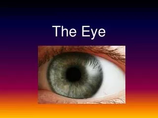
The eyeball is a slightly irregular hollow sphere with anterior and posterior poles, composed of three tunics: fibrous, vascular, and sensory. The outermost fibrous tunic includes the opaque sclera and the clear cornea, which protects the eye and allows light entry. The vascular tunic (uvea) consists of the choroid, ciliary body, and iris, regulating light through the pupil. The sensory tunic, or retina, contains photoreceptors that convert light into neural signals. The eye's structure supports vision and light regulation, highlighting the complex relationship between its components.

The Eye
E N D
Presentation Transcript
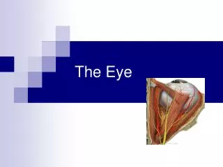
Structure of the Eyeball • Slightly irregular hollow sphere with anterior and posterior poles • The wall is composed of three tunics- fibrous, vascular and sensory • The internal cavity is filled with fluids called humors. • The lens separates the internal cavity into anterior and posterior segments
Fibrous Tunic • Forms the outermost coat of the eye: • Opaque sclera (posterior) • Clear cornea (anterior) • Sclera protects the eye • Cornea lets light enter the eye
Vascular Tunic (Uvea): Choroid Region • Has three regions: • Choroid • Cilliary body • Iris • Choroid Region: • A dark brown membrane that forms the posterior portion of the uvea • Supplied blood to all eye tunics
Vascular Tunic: Cilliary Body • Thickened ring of tissue surrounding the lens • Composed of smooth muscle bundles • Anchors the suspensory ligament that holds the lens in place
Vascular Tunic: Iris • The colored part of the eye • Pupil- central opening of the iris • Regulates amount of light entering the eye during: • 1. close vision and bright light- pupil contracts • 2. distant vision and dim light- pupil dilates • 3. changes in emotional state- pupils dilate when the subject matter is appealing or requires problem solving skills
Sensory Tunic: Retina • A delicate two-layered membrane • Pigmented layer- the outer layer absorbs light and prevents scattering • Neural Layer, which contains: • Photoreceptors that transduce light energy • Bipolar cells and ganglion cells • Amacrine and horizontal cells
The Retina: Ganglion cells and the Optic Disc • Ganglion cell axons: • Run along the inner surface of the retina • Leave the eye as the optic nerve • The optic disc: • Is the site where the optic nerve leaves the eye • Lacks photoreceptors (blind spot)
The Retina: Photoreceptors • Rods: • Respond to dim light • Are used for peripheral vision • Cones: • Respond to bright light • Have high-acuity color vision • Are found in the macula lutea • Are concentrated in the fovea centralis
Blood Supply to the Retina • The neural retina receives its blood supply from two sources • The outer third receives its blood from the choroid • The inner two-thirds are served by the central artery and vein • Small vessels radiate out from the optic disc and can be seen with an ophthalmoscope.