Understanding the Structure and Function of the Eye
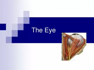
The eye is a complex organ essential for vision, likened to a digital camera. It measures around 25 mm in diameter and is supported by six extrinsic muscles within a protective bony orbit. The eye comprises three main layers: the corneoscleral coat (outer layer), uvea (middle layer), and retina (inner layer). The cornea, sclera, uvea, and retina work together to focus light, stabilize vision, and transmit visual information to the brain. Each structure within the eye performs vital functions for clarity and protection.


Understanding the Structure and Function of the Eye
E N D
Presentation Transcript
The eye Dr. MakarchukIryna
The eye is a complex sensory organ that provides the senseof sight. In many ways, the eye is similar to a digital camera. GENERAL STRUCTURE OF THE EYE • The eye measures approximately 25 mm in diameter. It is suspended in the bony orbital socket by six extrinsic muscles that control its movement. • A thick layer of adipose tissue partially surrounds and cushions the eye as it moves within the orbit. • The extraocular muscles are coordinated so that the eyes move symmetrically around their own central axes.
Layers of the Eye The wall of the eye consists of three concentric layers or coats: • The corneoscleral coat, the outer or fibrous layer, includes the sclera, the white portion, and the cornea, the transparent portion. • The vascular coat, the middle layer, or uvea, includes the choroid and the stroma of the ciliary body and iris. • The retina, the inner layer, includes an outer pigment epithelium, the inner neural retina, and the epithelium of the ciliary body and iris. The neural retina is continuous with the central nervous system through the optic nerve.
The corneoscleral coat consists of the transparent cornea and the white opaque sclera. • The cornea covers the anterior one-sixth of the eye. In this windowlike region, the surface of the eye has a prominence or convexity. • The cornea is continuous with the sclera. The sclera is composed of dense fibrous connective tissue that provides attachment for the extrinsic muscles of the eye. The sclera constitutes the “white” of the eye. In children, it has a slightly blue tint because of its thinness; in elderly people, it is yellowish because of the accumulation of lipofuscin in its stromal cells. • The corneoscleral coat encloses the inner two layers except where it is penetrated by the optic nerve.
The uvea consists principally of the choroid, the vascularlayer that provides nutrients to the retina. • Blood vessels and melanin pigment give the choroid an intense dark brown color. The pigment absorbs scattered and reflected light to minimize glare within the eye. • The anterior rim of the uveal layer continues forward, where it forms the stroma of the ciliary body and iris. The ciliary body is a ringlike thickening that extends inward just posterior to the level of the corneoscleral junction. Within the ciliary body is the ciliary muscle, a smooth muscle that is responsible for lens accommodation. Contraction of the ciliary muscle changes the shape of the lens, which enables it to bring light rays from different distances to focus on the retina. • The iris is a contractile diaphragm that extends over the anterior surface of the lens. It also contains smooth muscle and melanin-containing pigment cells scattered in the connective tissue.
The retina consists of two components: The neural retinaand pigment epithelium. The retina is a thin, delicate layer consisting of two components: • The neural retina is an inner layer that contains light sensitive receptors and complex neuronal networks. • The retinal pigment epithelium (RPE) is an outer layer composed of simple cuboidal melanin-containing cells. Externally, the retina rests on the choroid; internally, it is associated with the vitreous body. The neural retina consists largely of photoreceptor cells, called retinal rods and cones, and interneurons. Visual information encoded by the rods and cones is sent to the brain via impulses conveyed along the optic nerve.
Chambers of the Eye The layers of the eye and the lens serve as boundaries for three chambers within the eye. The chambers of the eye are the following: • The anterior chamber is the space between the cornea and the iris. • The posterior chamber is the space between the posterior surface of the iris and the anterior surface of the lens. • The vitreous chamber is the space between the posterior surface of the lens and the neural retina (Fig. 24.2). The cornea, the anterior and posterior chambers, and their contents constitute the anterior segment of the eye. The vitreous chamber, visual retina, RPE, posterior sclera, and uvea constitute the posterior segment.
MICROSCOPIC STRUCTURE OF THE EYECorneoscleral Coat The cornea consists of five layers: Three cellular layers and two noncellular layers. • The transparent cornea is only 0.5 mm thick at its center and about 1 mm thick peripherally. It consists of three cellular layers that are distinct in both appearance and origin. These layers are separated by two important membranes that appear homogeneous when viewed in the light microscope. Thus, the five layers of the cornea seen in a transverse section are the following: • Corneal epithelium • Bowman’s membrane (anterior basement membrane) • Corneal stroma • Descemet’s membrane (posterior basement membrane) • Corneal endothelium
Vascular Coat (Uvea) • The iris, the most anterior part of the vascular coat, forms a contractile diaphragm in front of the lens. • The sphincter pupillae is innervated by parasympathetic nerves; the dilator pupillae muscle is under sympathetic nerve control. • The ciliary body is the thickened anterior portion of the vascular coat and is located between the iris and choroid. • The ciliary muscle is organized into three functional portions or groups of smooth muscle fibers. • Ciliary processes are ridgelike extensions of the ciliary body from which zonular fibers emerge and extend to the lens. The processes and the ciliary body are covered by a double layer of columnar epithelial cells, the ciliary epithelium, which was originally derived from the two layers of the optic cup. The ciliary epithelium has three principal functions: • Secretion of aqueous humor • Participation in the blood–aqueous barrier (part of the blood–ocular barrier • Secretion and anchoring of the zonular fibers that form the suspensory ligament of the lens
The choroid is the portion of the vascular coat that lies deep to the retina. • The choroid is a dark brown vascular sheet only 0.25 mm thick posteriorly and 0.1 mm thick anteriorly. It lies between the sclera and retina. Two layers can be identified in the choroid: • The choriocapillary layer, an inner vascular layer • Bruch’s membrane, a thin, amorphous hyaline membrane
Retina • The retina represents the innermost layer of the eye. • The retina, derived from the inner and outer layers of the optic cup, is the innermost of the three concentric layers of the eye. It consists of two basic layers: • The neural retina or retina proper is the inner layer that contains the photoreceptor cells. • The RPE is the outer layer that rests on and is firmly attached through the Bruch’s membrane to the choriocapillary layer of the choroid.
In the neural retina, two regions or portions that differ in function are recognized: • The nonphotosensitive region (nonvisual part), located anterior to the oraserrata, lines the inner aspect of the ciliary body and the posterior surface of the iris (this portion of the retina is described in the sections on the iris and ciliary body). • The photosensitive region (optic part) lines the inner surface of the eye posterior to the oraserrata except where it is pierced by the optic nerve.
Layers of the Retina Ten layers of cells and their processes constitute the retina. Before discussing the ten layers of the retina, it is important to identify the types of cells found there. This identification will aid in understanding the functional relationships of the cells. For convenience, neurons and supporting cells can be classified into four groups of cells: • Photoreceptor cells — the retinal rods and cones • Conducting neurons — bipolar neurons and ganglion cells • Association neurons and others — horizontal, centrifugal, interplexiform, and amacrine neurons • Supporting (neuroglial) cells — Müller’s cells, microglial cells, and astrocytes
The specific arrangement and associations of the nuclei and processes of these cells result in the retina being organized in ten layers that are seen with the light microscope. The ten layers of the retina, from outside inward, are: 1. Retinal pigment epithelium (RPE)—the outer layer of the retina, actually not part of the neural retina but intimately associated with it 2. Layer of rods and cones—contains the outer and inner segments of photoreceptor cells 3. Outer limiting membrane—the apical boundary of Müller’s cells 4. Outer nuclear layer—contains the cell bodies (nuclei) of retinal rods and cones 5. Outer plexiform layer—contains the processes of retinal rods and cones and processes of the horizontal, amacrine, and bipolar cells that connect to them 6. Inner nuclear layer—contains the cell bodies (nuclei) of horizontal, amacrine, bipolar, and Müller’s cells 7. Inner plexiform layer—contains the processes of horizontal, amacrine, bipolar, and ganglion cells that connect to each other 8. Ganglion cell layer—contains the cell bodies (nuclei) of ganglion cells 9. Layer of optic nerve fibers—contains processes of ganglion cells that lead from the retina to the brain 10. Inner limiting membrane—composed of the basal lamina of Müller’s cells
Vision is a process by which light striking the retina is converted into electrical impulses that are transmitted to the brain. The impulses produced by light reaching the photoreceptor cells are conveyed to the brain by an elaborate network of nerves. The conversion of the incident light into nerve impulses is called visual processing and involves two basic steps: • Step 1 is a photochemical reaction that occurs in the outer segment of the rod and cone receptors as absorbed light energy causes conformational changes in the chromophores. • Step 2 consists of a decrease in the concentration of cGMP within the cytoplasm of the inner segment of the photoreceptor cells.
Crystalline Lens The lens is a transparent, avascular, biconvex structure. It is suspended between the edges of the ciliary body by the zonular fibers. The pull of the zonular fibers keeps the lens in a flattened condition. Release of tension causes the lens to fatten or accommodate to bend light rays originating close to the eye so that they focus on the retina. The lens has three principal components: • The lens capsule is a thick basal lamina measuring approximately 10 µm to 20 µm produced by the anterior lens cells. • The Subcapsular epithelium is a cuboidal layer of cells present only on the anterior surface of the lens. • The lens fibers are structures derived from subcapsular epithelial cells.
Changes in the lens are associated with aging. With increasing age, the lens gradually loses its elasticity and ability to accommodate. This condition, called presbyopia, usually occurs in the fourth decade of life. It is easily corrected by wearing reading glasses or using a magnifying lens. Loss of transparency of the lens or its capsule is also a relatively common condition associated with aging. This condition, called cataract, may be caused by conformational changes or cross-linking of proteins. The development of a cataract may also be related to disease processes, metabolic or hereditary conditions, trauma, or exposure to a deleterious agent (such as ultraviolet radiation). Cataracts that significantly impair vision can usually be corrected surgically by removing the lens and replacing it with a plastic lens implanted in the posterior chamber.
Vitreous Body • The vitreous body is the transparent jellylike substance that fills the vitreous chamber in the posterior segment of the eye. • The vitreous body is loosely attached to the surrounding structures, including the inner limiting membrane of the retina. • The main portion of the vitreous body is a homogeneous gel containing approximately 99% water (the vitreous humor), collagen, glycosaminoglycans (principally hyaluronan), and a small population of cells called hyalocytes. These cells are believed to be responsible for synthesis of collagen fibrils and glycosaminoglycans.
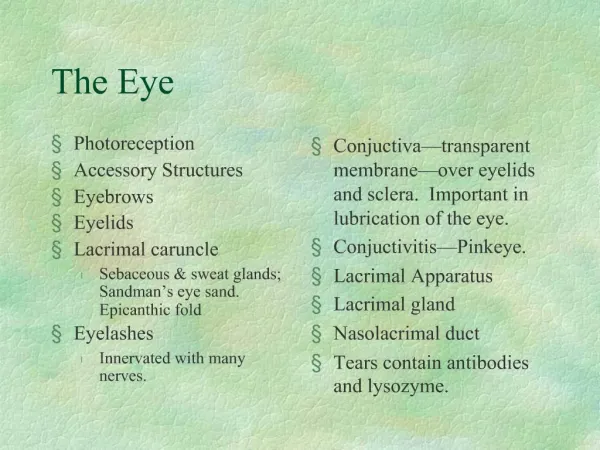
Accessory Structures of the Eye • The conjunctiva lines the space between the inner surface of the eyelids and the anterior surface of the eye lateral to the cornea. • The conjunctiva is a thin, transparent mucous membrane that extends from the corneosclerallimbus located on the peripheral margin of the cornea across the sclera (bulbar conjunctiva) and covers the internal surface of the eyelids (palpebral conjunctiva). It consists of a stratified columnar epithelium containing numerous goblet cells and rests on a lamina propria composed of loose connective tissue. The goblet cell secretion is a component of the tears that bathe the eye. • Conjunctivitis, an inflammation of the conjunctiva that is commonly called pinkeye, is characterized by redness, irritation, and watering of the eyes.
The primary function of the eyelids is to protect the eye. • The skin of the eyelids is loose and elastic to accommodate their movement. Within each eyelid is a flexible support, the tarsal plate, consisting of dense fibrous and elastic tissue. • In addition to eccrine sweat glands, which discharge their secretions directly onto the skin, the eyelid contains four other major types of glands: • The tarsal glands (Meibomian glands), long sebaceous glands embedded in the tarsal plates, appear as vertical yellow streaks in the tissue deep in the conjunctiva. • Sebaceous glands of eyelashes (glands of Zeis) are small, modified sebaceous glands that are connected with and empty their secretion into the follicles of the eyelashes. • Apocrine glands of eyelashes (glands of Moll) are small sweat glands with unbranched sinuous tubules that begin as a simple spiral. • Accessory lacrimal glands are compound serous tubuloalveolar glands that have distended lumina. They are located on the inner surface of the upper eyelids (glands of Wolfring) and in the fornix of the lacrimal sac (glands of Krause).
The lacrimal gland produces tears thatmoisten the corneaand pass to the nasolacrimalduct • Tears are produced by the lacrimal glands and to a lesser degree by the accessory lacrimal glands. The lacrimal gland islocated beneath the conjunctiva on the upper lateral side of the orbit. The lacrimal gland consists of several separate lobules of tubuloacinar serous glands. • Tears keep the conjunctiva and corneal epithelium moist and wash foreign material from the eye as they flow across the corneal surface toward the medial angle of the eye.