Spectroscopy Workshop
Spectroscopy Workshop. School of Chemistry The Queen’s University of Belfast. Workshop Content. Spectroscopy overview Ultra-violet/visible (UV-vis) Infra-Red (IR) Nuclear Magnetic Resonance (NMR) Mass Spectrometry. Spectroscopy.

Spectroscopy Workshop
E N D
Presentation Transcript
Spectroscopy Workshop School of Chemistry The Queen’s University of Belfast
Workshop Content Spectroscopy overview Ultra-violet/visible (UV-vis) Infra-Red (IR) Nuclear Magnetic Resonance (NMR) Mass Spectrometry
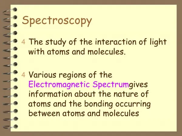
Spectroscopy In spectroscopy, transitions between different energy levels within atoms and molecules are recorded and then used to give information on chemical structure.
Visible X-Rays Microwave UV IR Gamma Rays Radio 10- 11 10- 9 10- 7 10- 5 10- 3 10- 1 10 10 3 Wavelength (cm) The range of energies that can be used for spectroscopy is very large and spans a large proportion of the electromagnetic spectrum.
After Absorption DE Energy In a typical experiment, the molecules or atoms start at lower energy and go to a higher energy level upon absorption of radiation of appropriate wavelength. Before Energy
After After Energy Absorption can only occur when the energy of the radiation (calculated from the frequency or wavelength) matches the energy gap. If there are several different upper levels (and there usually are) then several transitions will be observed. After Before
For current purposes we look only at: UV/visible ( highest energy) Infra red (intermediate) Radio frequency (lowest energy). But in all cases :
To record a spectrum, sweep through the appropriate range of energies and look for absorption at particular values.
Absorption gives peaks, when these have been measured this gives the energy gaps within the sample. These can then be related to structure. Interpretation depends on the energy range investigated.
UV/visible Spectroscopy Chemical compounds are coloured because they absorb visible light. In general, even organic compounds that are colourless will absorb UV light.
Absorption of visible light Where has the energy that was within the photons gone to ?
After DE Energy In UV/visible spectroscopy the energy of the absorbed photon is used is used to drive the molecule into an excited electronic state. In the excitation the energy of the whole molecule increases. Before Absorption Energy
This overall change is typically due to promotion of a single electron from a lower to higher energy orbital. The energy of the transition depends on the gap between the two orbitals. In organic compounds which have only single bonds between the atoms the excitation energy is very high- lies in deep UV.
a bonding to an anti-bonding orbital. e.g. ethene Even if have a simple p bond, the excitation from highest occupied to lowest unoccupied orbitals still lies in the UV. This excitation gives a dramatic decrease in bond order due to excitation from
If we have a highly conjugated molecule the energy separation between the orbitals is smaller. Excitation of the electron thus has a proportionately smaller effect and requires less energy- energy gap may lie in the visible region.
Orbitals of Butadiene Anti-bonding Energy Bonding Again note that lowest energy transition may lie in visible. But we can also excite to higher orbitals with sufficiently energetic (UV) photons.
With increasing conjugation, the decreasing energy gap is reflected by absorption at longer wavelengths.
beta-Carotene trans-Crocetin Xanthopterin 16,17-DimethoxyViolanthrone The structures of many coloured compounds show they are very extensively conjugated.
PURPLE ORANGE BLUE Substituents added to the compound may alter the energy of the orbitals which e- is excited from or to. Auxochromes: substituents that alter the wavelength or intensity of the absorption due to the chromophore
-2H+ HO HO O- O- pink colourless Changes in chemical composition can give rise to pronounced colour changes since this can dramatically alter the energies of the orbitals involved in the transitions e.g. pH indicators. Phenolphthalein
red H+ orange-yellow Methylorange
Summary Absorption of UV-vis radiation occurs via excitation of electrons from filled to unfilled orbitals i.e. they are electronic transitions. Molecules have characteristic absorption spectra. The absorption can lead to coloured materials. pH Indicators use the change in colour between the acid and alkali forms of the molecules.
IR Spectroscopy Origin of the absorption The spectrometer The spectra Organic compounds Example problem
Origin of IR absorptions CO2 symmetric stretch Atoms within a molecule are never still. They vibrate in a variety of ways (modes). Atoms may be considered as weights connected by springs. Each vibrational mode has its own resonant frequency. asymmetric stretch bending
If the vibrational mode involves a change in molecular dipole moment, the vibration can be induced by absorption of a photon - it is ‘IR-active’ Appropriate energy for this is infra-red symmetric stretch nodipole nodipole asymmetric stretch change in dipole - IR active bending change in dipole - IR active
The bigger the change in dipole, the more intense the absorption 100 Transmittance /% 0 Stretching higher energy than bending 2800 2400 2000 1600 1200 800 400 CO2 IR spectra Wavenumber /cm-1 The symmetric stretch is not IR active (no change in dipole)
More complex: Wavenumber/cm-1 Ethyl ethanoate (CH3COOCH2CH3) 2000 3000 1500 1000 500 4000 C=O bond C-O stretch IR spectra of organic compounds
Wavenumber / cm- 1 4000 3000 2000 1500 1000 650 (all types) N C C O C Cl H O H H C C C C C N O C But functional groups have characteristic frequencies
Four regions in the spectrum: Wavenumber / cm-1
Example problem • Explain why infrared radiation is absorbed by molecule HCl but not by molecules H2 and Cl2. • Explain what occurs in the HCl molecule when infrared radiation is absorbed. • The simplified infrared spectrum below is that of an organic compound. 100 90 80 Transmittance / % 70 60 50 40 30 20 10 4000 3600 3200 2800 2400 2000 1900 1800 1700 1600 Wavenumber / cm-1 • Identify two main functional groups on the spectrum. • This compound has composition by mass C, 67.9%; H, 5.7%; N, 26.4%, and Mr of 53. Suggest a structural formula for the compound. C=C C=O? C=N? C-H CC CN? Identify two main functional groups present in the compound which gave this spectrum
Combine this information with the following data to deduce its structure C 67.9% H 5.7% N 26.4% Mr 53 Likely structure: C Cyanoethene So, formula = C3H3N
Absorption of IR can occur if a vibrational mode is associated with a change in dipole. Functional groups have characteristic absorption frequencies. In combination with other analytical data, the structure of an organic compound can often be deduced. Summary
NMR Spectroscopy The Basis of NMR Spectroscopy The Spectrometer Chemical Shifts Signal Intensity and Integration Coupling Constants Example Spectra
The Basis of NMR Spectroscopy Atomic nuclei behave like small bar magnets as a result of their charge and spin. In the presence of an applied magnetic field the spin states have different energy and the magnetic moment can align with or against the applied field.
The difference in energy between the two spin states is dependent on the external magnetic field strength. Irradiation of a sample with radio frequency energy corresponding to the spin state separation (DE) will excite nuclei in the +½ state to the higher energy –½ state.
The 1H NMR Experiment For example, consider a water sample in a 2.3487 T external magnetic field irradiated by 100 MHz radiation. If the magnetic field is increase to 2.3488 T the water protons will at some point absorb rf energy (DE) and a resonance signal will appear,
The Chemical Shift Not all protons give resonance signals at the same field frequency. Electrons move in response to the applied field and generate a secondary magnetic field which opposes the applied field. The secondary field shields the nucleus from the applied field and nuclei in different environments resonate at different frequencies. The difference in resonance frequency is measured as a chemical shift, units d
MEK Signal Intensity The relative area of the absorption signals can provide valuable structural information. The area under a peak is proportional to the number of a given type of nuclei in the molecule.
The keto-enol equilibrium ratio of 2,4-pentandione can determined by 1H NMR spectroscopy
The applied magnetic field experienced by a proton Ha will be modified by the local field produced by its neighbouring Hb Ha modifies the field at Hb by aligning with or against the applied field and and gives 2 resonant frequencies for Hb (doublet) Spin-Spin Coupling Similarly Hb modifies the field at Ha in 3 different ways (triplet)
Splitting pattern can provide valuable structural information Chemically equivalent protons act as a group and a peak due to n adjacent protons is split into n+1 lines, with a coupling constant J
1H NMR Spectrum of Ethyl Acetate
1H NMR Spectrum of 1,3-Dichloropropane
Example Problem Given the formula, deduce what you can about the structure Integration corresponds to 2H : 2H : 3H A triplet must correspond to 2 near neighbour protons A sextet corresponds to 5 near neighbour protons Therefore CH2, CH2 and CH3 groups are present
Connectivity can be deduced to be Solution
Summary NMR spectroscopy involves irradiating a sample with radio frequency radiation Protons in different chemical environments have different chemical shifts d Protons in different environments can couple to each other with a coupling constant J The combination of chemical shifts and coupling constants provides valuable structural information
Mass Spectrometry • The basic principles • Applications
What is a mass spectrometer ? • A mass spectrometer is an instrument which produces charged particles (ions) from chemical substances under analysis. • It then uses magnetic and/or electric fields to separate those ions and to measure their mass.