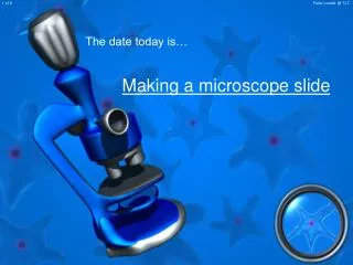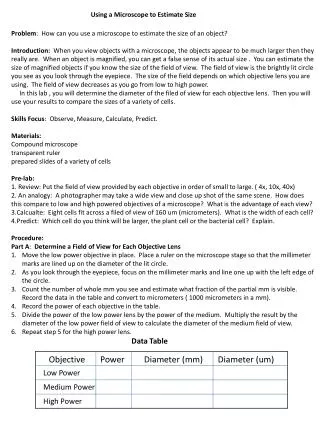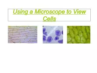
Using a Microscope
Using a Microscope. Parts of a Microscope. Field of View. As magnification increases, the field of view decreases!. At 40x magnification you will be able to see 5mm. At 100x magnification you will be able to see 2mm. At 400x magnification you will be able to see 0.45mm, or 450 microns.


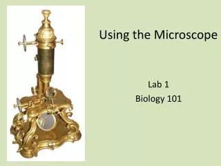
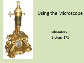
Using a Microscope
E N D
Presentation Transcript
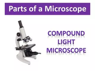
Parts of a Microscope
Field of View As magnification increases, the field of view decreases!
At 40x magnification you will be able to see 5mm. • At 100x magnification you will be able to see 2mm. • At 400x magnification you will be able to see 0.45mm, or 450 microns. • At 1000x magnification you will be able to see 0.180mm, or 180 microns.
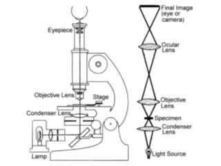
Focusing Microscopes Because specimens are three dimensional, and vary in thickness, you must make adjustments as you view the specimen. This is especially true if you are looking a live organisms that are moving.
Calculating Magnification The magnification of an object is calculated by multiplying the magnification of the eyepiece lens (10X) by the magnification of the objective lens. Ex) 10X x 43X = 430X
10X 20X 40X
Using A Microscope • Start on the low objective lens with the stage lowered and the specimen placed on the stage. • Adjust the course adjustment knob until you can see the image. • Use the fine adjustment knob to focus clearly on the specimen. • Rotate the nosepiece to high power. Do not move the stage! • Use only the fine adjustment knob to bring the specimen into focus!
Escherica coli (bacterium) 1000X .5 - .8 um
Human Red Blood Cell 1000X 6 - 8 um
Human Egg Cell 1000X 100 um
Saccharomyces cerevisiae(Yeast) 1000X 5 - 10 um
Streptococcus pneumoniae(Bacterium) 1000X .5 – 1.3 um