Mastering Microscopy Techniques for Enhanced Image Resolution
Learn how to adjust resolution, contrast, and brightness with a microscope, from sample preparation to image enhancement. Explore light microscopy modifications and electron microscopy variations to optimize sample analysis.

Mastering Microscopy Techniques for Enhanced Image Resolution
E N D
Presentation Transcript
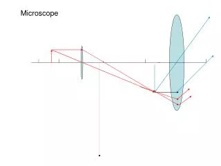
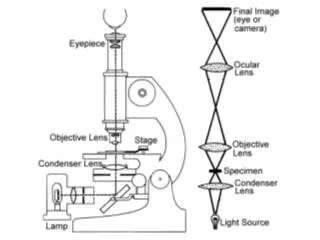
resolution contrast brightness Adjusting a Microscope • Center components on optic axis • Focus objective • Focus condenser • Adjust illumination • lamp voltage (intensity) • iris diaphragm
Sample Preparation • fixation • organic solvents (acetone, alcohols) • formaldehyde • glutaraldehyde • sectioning • for tissues or thick samples • embed in parrafin or resin • cut with microtome • staining • dyes differentially bind to DNA, RNA and protein • provides more contrast
Light Microscopy Modifications • Phase Contrast • Differential Interference Contrast (Normarski) • Confocal Scanning • Fluorescence • Dark Field (diffracted light) • Image Enhancement
Phase Contrast and Differential Interference Contrast • requires special objective and condenser lens • phase differences are converted into intensity differences • distinguish objects that only differ slightly in refractive index or thickness
Light Microscopy Modifications • Phase Contrast • Differential Interference Contrast (Normarski) • Confocal Scanning • Fluorescence • Dark Field (diffracted light) • Image Enhancement
resolution contrast brightness Image Enhancement • video cameras + computers used to enhance images • correct imperfections in optical systems • overcome limitations of human eye • seeing image in dim light • seeing small intensity differences against bright background • does not increase actual resolution
Limit of Resolution • distance at which two objects can be resolved • resolution limit = 0.61/numerical aperture • (NA is a lens property) • of visible light = 0.4-0.8 mm • Electron Microscopy • samples are analyzed with electrons • particles traveling near the speed of light behave as a wave • wavelength with velocity • resolutions of 2 nm or less
Sample Preparation • Fixation • glutaraldehyde • osmium tetroxide • Dehydration • ethanol (step-wise) • Embedding • plastic resins • Sectioning • ultramicrotome (50-100 nm thick) • Staining • heavy metals
Variations of Electron Microscopy • Transmission (TEM) • Scanning (SEM) • Shadow-casting • Freeze-fracture • Freeze-etching • CryoEM • Negative Staining