Download

1 / 20
200 likes | 302 Vues
Explore the composition and structure of bone tissue including organic and inorganic matter. Learn about types of bone cells and the control of calcium concentration in blood. Discover the anatomy and features of long bones.

E N D
BONE HISTOLOGY Bone Histology Lab # 3
BONE HISTOLOGY (Composition) A- Organic matter COMPOSITION OF BONE TISSUE A- Organic matter. B- Inorganic matter. Cells: Osteoblasts,osteocytes,osteoclasts,and osteoprogenitor cells. Collagen fibers : Provides tensile strength. Polysaccharides : Help to form the ground substance of bone.
BONE HISTOLOGY (Composition) Types of Bone Cells 1- Osteoprogenitor cell: Stem cell whose divisions produce the osteoblasts. 2- Osteoblast: Immature bone cell that secrets organic components of the matrix. 3- Osteocyte: Mature bone cell that maintains the bone matrix. 4- Osteoclast: Multinucleated cell that secrets acids and enzymes to dissolve bone matrix.
BONE HISTOLOGY (Composition) Types of Bone Cells
Osteoclasts dissolve bone here Osteoblastsadd bone by appositional growth Remodeling Growth
CONTROL OF CALCIUM CONCENTRATION IN BLOOD Parathyroid hormone is released by parathyroid gland Stimulates the osteoclaststo resorb bone, releasing calcium to the blood [Calcium] in blood Calcitonin hormone is released by thyroid gland Inhibits osteoclasts to resorb bone, and encourages calcium salt deposit in bone matrix [Calcium] in blood First Hormonal Mechanism [Calcium] in blood Second Hormonal Mechanism [Calcium] in blood
BONE HISTOLOGY (Composition) B- Inorganic matter Hydroxyapatite: A combination of calcium and phosphorus salts that make the bone hard Minerals: Mainly magnesium and sodium ions that help form matrix
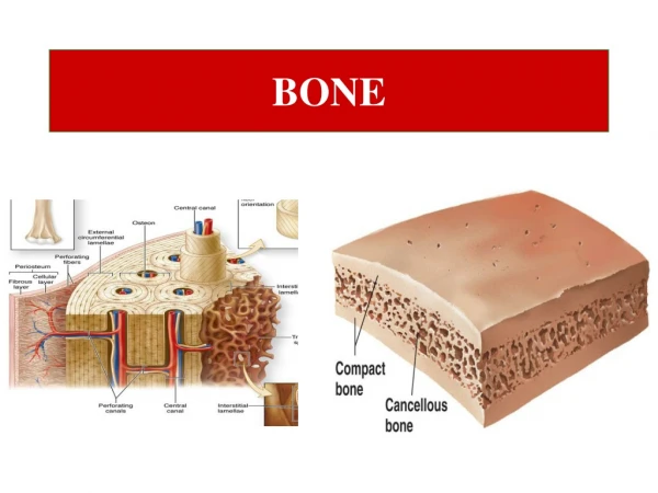
Epiphysis Metaphysis Compact bone Spongy bone Metaphysis Epiphysis STRUCTURE OF BONE Spongy bone Compact bone Marrow cavity Diaphysis
A. Long Bone Anatomy Proximal epiphysis Articular cartilage Epiphyseal plate Diaphysis Endosteum Osteon Spongy bone Periosteum Sharpey’s fibers Red bone marrow Marrow or Medullary cavity with yellow bone marrow Distal epiphysis Compact bone
Compact Bone Tissue It is the structural and functional unit of the compact bone Osteon Concentric lamellae External circumferential lamellae Interstitial lamellae Periosteum Trabeculae of spongy bone Perforating (Sharpey’s) fibers They contain blood capillaries, lymphatic vessels, and nerves Central (Harversian) canals They connect the central canals of adjacent osteons to each other Perforating (Volkmann’s) canals with blood vessels
Compact Bone Tissue Osteocyte in lacuna They are bone maintaining cells Interstitial lamellae Concentric lamellae Central (Harversian) canal Artery Vein Nerve
Compact Bone Tissue They contain the osteocytes In Central (Harversian) canal Lacunae Artery Nerve Interstitial lamellae Vein Concentric lamellae They allow delivery of nutrients and removal of waste products to and from the osteocytes enclosed in the hard matrix Osteocytes in lacunae Canaliculi Structure of the Osteon
BONE HISTOLOGY (Structure)
Skeleton Axial Skeleton Appendicular Skeleton
Head of the femur Head of the humerus Costal facets Condyles Anatomical Features (markings) of Bones Projections that help to form joints: Olecranon process Process: Any bony prominence
Trochanters Tibial tuberosity Linea aspera Anterior crest Epicondyle Condyle Lesser tubercle Spine of scapula Projections that are sites of muscle and ligament attachment:
Fovea capitis Frontal sinus Internal acoustic meatus Hypophyseal Fossa Infraorbital foramen Superior orbital fissure Depressions: Alveolus: A pit or socket (tooth socket) Alveolus Fovea: A small pit Passages and cavities: Canal: A tubular passage or tunnel in a bone Meatus: An opening into a canal
The Shapes of Bones • Irregular bones • Flat bones • They have elaborate shapes that don’t fit into the other categories. • They protect soft organs. They are • curved but wide & thin. • Long bones • Short bones • Longer than wide. They serve as rigid levers that are acted upon by skeletal muscles to produce body movements. • They are equal in length and width. • They glide across one another in multiple directions. • Sutural bones • Sesamoid bones (patella)