Colorectal cancer
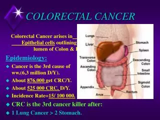
Colorectal cancer. Rowa ’ Al- Ramahi. PATHOPHYSIOLOGY. Development of a colorectal neoplasm is a multistep process of genetic and phenotypic alterations of normal bowel epithelium structure and function leading to unregulated cell growth, proliferation, and tumor development.

Colorectal cancer
E N D
Presentation Transcript
Colorectal cancer Rowa’ Al-Ramahi
PATHOPHYSIOLOGY • Development of a colorectal neoplasm is a multistep process of genetic and phenotypic alterations of normal bowel epithelium structure and function leading to unregulated cell growth, proliferation, and tumor development.
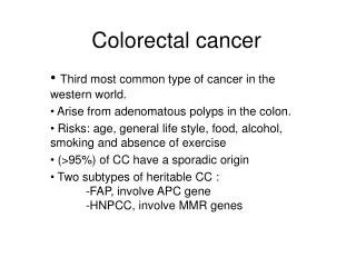
Features of colorectal tumorigenesisinclude genomic instability, activation of oncogene pathways, mutational inactivation of tumor-suppressor genes, and activation of growth factor pathways. • Adenocarcinomas account for more than 90% of tumors of the large intestine.
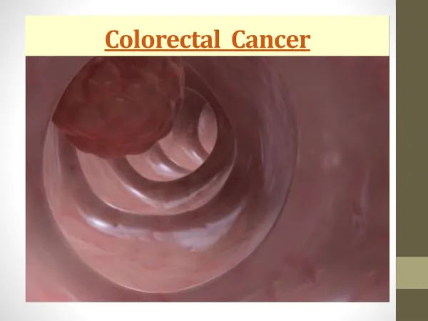
CLINICAL MANIFESTATIONS • Signs and symptoms of colorectal cancer can be extremely varied, subtle, and nonspecific. Patients with early-stage colorectal cancer are often asymptomatic, and lesions are usually detected by screening procedures. • Blood in the stool is the most common sign; however, any change in bowel habits, vague abdominal discomfort, or abdominal distension may be a warning sign.
Less common signs and symptoms include nausea, vomiting, and, if anemia is severe, fatigue. • Approximately 19% of patients with colorectal cancer present with metastatic disease. The most common site of metastasis is the liver, followed by the lungs, and then bones.
PREVENTION AND SCREENING • Primary prevention is aimed at preventing colorectal cancer in an at-risk population. To date, the only strategy shown to reduce the risk is chemoprevention with celecoxib in people with familial adenomatouspolyposis. • Secondary prevention is aimed at preventing malignancy in a population that has already manifested an initial disease process.
Secondary prevention includes procedures ranging from colonoscopic removal of precancerous polyps detected during screening colonoscopy to total colectomy for high-risk individuals (e.g., familial adenomatouspolyposis). • Current US guidelines for average-risk individuals include annual occult fecal blood testing starting at age 50 years and examination of the colon every 5 or 10 years depending on the procedure.
DIAGNOSIS • When colorectal carcinoma is suspected, a careful personal and family history and physical examination should be performed. • The entire large bowel should be evaluated by colonoscopy or flexible sigmoidoscopy with double-contrast barium enema.
Baseline laboratory tests should include complete blood cell count, INR, activated partial thromboplastin time, liver and renal function tests, and serum carcinoembryonic antigen (CEA). Serum CEA can serve as a marker for monitoring colorectal cancer response to treatment, but it is too insensitive and nonspecific to be used as a screening test for early-stage colorectal cancer.
Radiographic imaging studies may include chest x-ray, bone scan, chest and abdominal computed tomography scans, positron emission tomography, ultrasonography, and magnetic resonance imaging. • Immunodetection of tumors using tumor-directed antibodies is an imaging technique for determining the location and extent of extrahepatic disease. These tests might also be useful for identifying metastatic or recurrent disease in patients with rising CEA levels.
Stage of colorectal cancer should be determined at diagnosis to predict prognosis and to develop treatment options. Stage is based on the size of the primary tumor (T1–4), presence and extent of lymph node involvement (N0–2), and presence or absence of distant metastase (M). • ✓ Stage I disease involves tumor invasion of the submucosa (T1) or muscularispropria (T2) and negative lymph nodes.
✓ Stage II disease involves tumor invasion through the muscularispropriainto pericolorectaltissues (T3), or penetration to the surface of the visceral peritoneum (T4a), or directly invades or is adherent to other organs or structures (T4b), and negative lymph nodes. • ✓ Stage III disease includes T1–4 AND positive regional lymph nodes. • ✓ Stage IV disease includes any T, any N, AND distant metastasis.
PROGNOSIS • Stage at diagnosis is the most important independent prognostic factor for survival and disease recurrence. Five year relative survival is ~92% for those with localized tumor at diagnosis as compared with 11% for those with metastatic disease at diagnosis.
PROGNOSIS • Poor prognostic clinical factors at diagnosis include bowel obstruction or perforation, rectal bleeding, high preoperative CEA level, distant metastases, and location of the primary tumor in the rectum or rectosigmoidarea. Studies to determine the prognostic use of genetic factors are ongoing.
DESIRED OUTCOME • The goal of treatment depends on the stage of disease. Stages I, II, and III are potentially curable; the intent is to eradicate micrometastatic disease. • Twenty to thirty percent of patients with metastatic disease may be cured if their metastases are resectable. Most stage IV disease is incurable; palliative treatment is given to reduce symptoms, avoid disease-related complications, and prolong survival.
TREATMENT • GENERAL PRINCIPLES • Treatment modalities are surgery, radiation therapy (RT), and chemotherapy and biomodulators. Adjuvant therapy for early stage disease and treatment of metastatic disease will be discussed separately.
SURGERY • Complete surgical resection of the primary tumor is the treatment of choice for most patients with operable disease. • Surgery for colon cancer generally involves complete tumor resection with an appropriate margin of tumor-free bowel and a regional lymphadenectomy.
Surgery for rectal cancer depends on the area involved. Although less than 33% of these patients require permanent colostomy, frequent complications include urinary retention, incontinence, impotence, and locoregional recurrence. • Common complications of surgery for both colon and rectal cancer include infection, anastomotic leakage, obstruction, adhesions, and malabsorption syndromes.
Adjuvant Therapy for Colon Cancer • Adjuvant therapy is administered after complete tumor resection to eliminate residual local or metastatic microscopic disease. Adjuvant therapy is not indicated for stage I colorectal cancer because >90% of patients are cured by surgical resection alone.
Adjuvant Therapy for Colon Cancer • Results of adjuvant chemotherapy studies in patients with stage II disease are conflicting. Despite a lack of consensus among practitioners, the approach to treatment of high-risk stage II and stage III disease is similar. • Adjuvant chemotherapy is the standard of care for stage III colon cancer.
Adjuvant Radiation Therapy • Adjuvant RThas no definitive role in colon cancer because most recurrences are usually extrapelvic & occur in the abdomen. • Acute adverse effects associated with RT include hematologic depression, dysuria, diarrhea, abdominal cramping, and proctitis. Chronic symptoms may persist for months after RT and may involve diarrhea, proctitis, enteritis, small-bowel obstruction, perineal tenderness, and impaired wound healing.
CHEMOTHERAPY • Fluorouracil (5-FU) is the most widely used chemotherapeutic agent for colorectal cancer. Leucovorin (folinic acid) is usually added to 5-FU as a biochemical modulator to enhance cytotoxic activity. • Administration method affects clinical activity and toxicity. 5-FU is administered by IV bolus or by continuous IV infusion. Efficacy evaluations favor continuous infusion 5-FU but none of the combination regimens with leucovorin has proven superior with regard to overall patient survival.
Continuous IV infusion of 5-FU is generally well tolerated but is associated with palmar-plantar erythrodysesthesia or hand–foot syndrome and stomatitis. IV bolus administration is associated with leukopenia, which is dose limiting and can be life threatening. Both methods are associated with a similar incidence of mucositis, diarrhea, nausea and vomiting, and alopecia.
In rare cases, patients deficient in dihydropyrimidinedehydrogenase, responsible for the catabolism of 5-FU, develop severe toxicity, including death, after 5-FU administration. • Capecitabine, an oral prodrug of 5-FU, has efficacy and safety profiles similar to those of IV infusion of 5-FU.
National guidelines recommend oxaliplatin-based regimens as the first line option for patients with stage III colon cancer who can tolerate combination therapy. It is commonly administered with fluorouracil/leucovorin. • Oxaliplatin is associated with paresthesia, neutropenia, and GI toxicity. Acute neuropathy is reversible within 2 weeks, usually occurs peripherally but may occur in the jaw and tongue, and is precipitated by exposure to cold. Persistent neuropathy is cumulative and is characterized by paresthesias, dysesthesias, and hypoesthesiasthat usually resolve with dosage reductions or cessation of oxaliplatin therapy.
Selection of an adjuvant regimen is based on patient-specific factors, including performance status, comorbid conditions, and patient preference based on lifestyle factors • Fluorouracil/leucovorin regimens currently have limited use but are acceptable options in patients who cannot receive oxaliplatin and are unable to tolerate oral capecitabine.
ADJUVANT THERAPY FOR RECTAL CANCER • Rectal cancer is more difficult to resect with wide margins, so local recurrences are more frequent than with colon cancer. Adjuvant RT plus chemotherapy is considered the standard of care for stages II and III rectal cancer.
RT reduces the risk of local tumor recurrence in patients undergoing surgery for rectal cancer. RT is given prior to surgery to decrease tumor size, making it more resectable. • Postoperative RT is used to treat a defined area but is associated with more toxicity.
Fluorouracil enhances the cytotoxic effects of RT. Compared with surgery alone, the combination of adjuvant fluorouracil and RT for 6 months reduces local and distant tumor recurrences and improves survival in stages II and III rectal cancer. • Preoperative (neoadjuvant) RT shrinks rectal tumors prior to surgical resection, improving sphincter preservation. Preoperative infusionalfluorouracilbasedchemotherapy plus RT is the preferred treatment for resectable T3 N0 or any T, N1–2 lesions. Patients should receive adjuvant chemotherapy following surgery to total 6 months of chemotherapy.
Neoadjuvant fluorouracil or capecitabinechemoradiationfollowed by surgery should be considered for locally unresectable tumors (T4). • All patients who receive preoperative chemotherapy for rectal cancer should receive postoperative chemotherapy, regardless of whether the disease was initially resectable.
METASTATIC DISEASE • Initial Therapy • Chemotherapy is the primary treatment modality for metastatic colorectal cancer (MCRC). Currently, most MCRCs are incurable. Initial chemotherapy is administered with palliative intent: to reduce symptoms, improve quality–of life, and extend survival. In general, treatment options are similar for metastatic cancer of the colon and rectum.
Surgical resection of discrete metastases in select patients may extend disease free survival. Resection of hepatic limited metastases may result in cure. Adjuvant chemotherapy may be administered, but the optimal regimen remains to be determined. • Neoadjuvantchemotherapy is administered to patients with liver metastases to increase complete resection rates with resectableor unresectableliver lesions. Oxaliplatinbased regimens with or without bevacizumabare commonly used. A total of 6 months of chemotherapy (preandpostoperative) should be administered. • Symptom control is the primary goal of RT in advanced or metastatic colorectal cancer.
Chemotherapy • Site(s) of tumor involvement and history of prior chemotherapy help define a management strategy for MCRC. Various chemotherapy regimens are recommended by national guidelines for initial palliative chemotherapy. The results of recent metaanalysessuggest that palliative chemotherapy improves survival in MCRC.
The most important factor in patient survival is not the initial chemotherapy regimen but ensuring that patients receive all three active drugs (fluorouracil, irinotecan, and oxaliplatin) at some point in their treatment course. Either irinotecanor oxaliplatinplus fluorouracil and leucovorinis recommended as first-line therapy for MCRC (FOLFOX, FOLFIRI). These regimens result in improved response rates and progression-free survival, as well as improved median survival. • Irinotecanis a topoisomerase I inhibitor. Early- and late-onset diarrhea and neutropenia are dose-limiting toxicities of irinotecan.
Early-onset diarrhea occurs 2 to 6 hours after administration and is characterized by lacrimation, diaphoresis, abdominal cramping, flushing, and/or diarrhea. These cholinergic symptoms respond t IV or subcutaneous atropine, 0.25 to 1 mg. • Late-onset diarrhea occurs 1 to 12 days after administration, lasts 3 to 5 days, and can be fatal. Late-onset diarrhea requires aggressive, high-dose loperamidebeginning with 4 mg after the first soft or watery stool, followed by 2 mg every 2 hours until symptom free for 12 hours. • Capecitabinemonotherapy is suitable for first-line therapy in patients not likely to tolerate IV chemotherapy.
Capecitabine is s suitable replacement for infusional fluorouracil in combination with oxaliplatin (CapeOx). • Bevacizumabis a humanized monoclonal antibody directed against vascular endothelial growth factor. The addition of bevacizumabto fluorouracil-based regimens improves response rates, time to progression, and median survival compared with chemotherapy alone, and the resulting four-drug regimens are considered firstlinetherapy for MCRC. • Bevacizumabis associated with hypertension, which is easily managed with oral antihypertensive agents. Other safety concerns are bleeding, thrombocytopenia, and proteinuria. GI perforation is a rare but potentially fatal complication necessitating prompt evaluation of abdominal pain associated with vomiting or constipation.
Cetuximabis a chimeric monoclonal antibody directed against epidermal growth factor receptor. Common adverse events include acne-like skin rash, asthenia, lethargy, malaise, and fatigue. • Treatment guidelines also include the addition of cetuximab to FOLFIRI, FOLFOX, or CapeOx as initial therapy in patients with wild-type KRAS tumors only.
Second-Line Therapy • The selection of second-line chemotherapy is primarily based on the type of prior therapy received, as well as the response to prior treatments, site and extent of disease, and patient factors and treatment preferences. The optimal sequence of regimens has not been established.
If disease progressed on first-line bevacizumab, data do not support continued use. • Cetuximab, either alone or in combination with irinotecan, can be used in patients with disease progression on irinotecan. Cetuximabmonotherapycan also be considered as salvage therapy in patients with irinotecan-or oxaliplatin-refractory disease. • Panitumumabis a human monoclonal antibody directed against epidermal growth factor receptor. Common adverse events are dermatologic toxicities, fatigue, abdominal pain, nausea, and diarrhea. As with cetuximab, the use of panitumumabshould be limited to patients with wildtypeKRAS tumors only.
Panitumumabis approved for use in MCRC that no longer responds to previous therapy with fluorouracil, irinotecan, or oxaliplatin. • Patients with hepatic-predominant disease whose disease progresses with systemic therapy may be candidates for hepatic-directed therapies such as chemoembolization, cryotherapy, or radiofrequency ablation.
EVALUATION OF THERAPEUTIC OUTCOMES • The goals of monitoring are to evaluate the benefit of treatment and to detect recurrence. • Patients who undergo curative surgical resection, with or without adjuvant therapy, require routine follow-up. • Patients should be evaluated for anticipated side effects such as loose stools or diarrhea, nausea or vomiting, mouth sores, fatigue, and fever. • Patients should be closely monitored for side effects that require aggressive intervention, such as irinotecan-induced diarrhea and beva-cizumab-induced GI perforation. Patients should be evaluated for other treatment-specific side effects, such as oxaliplatin-induced neuropathy, cetuximaband panitumumab-induced skin rash, and bevacizumab-induced hypertension and proteinuria.
EVALUATION OF THERAPEUTIC OUTCOMES • Less than one half of patients develop symptoms of recurrence, such as pain syndromes, changes in bowelhabits, rectal or vaginal bleeding, pelvic masses, anorexia, and weight loss. CEA levels may help detect recurrences in asymptomatic patients. • Quality-of-life indices should be monitored, especially in patients with metastatic disease.