Download

1 / 20
250 likes | 660 Vues
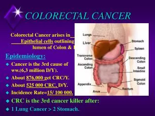
COLORECTAL CANCER . COLORECTAL CANCER. Incidence 2 nd after after bronhopulmonary C in males and breast in females >65 ani M :F= 1- 1,5:1. Food habits Excess of animal fat and colesterol Lack of fibers in food Excess of salt, spicy, smoked mea, alcohol, food additives Heredity

E N D
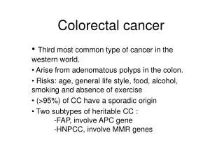
COLORECTAL CANCER • Incidence • 2nd after after bronhopulmonary C in males and breast in females • >65 ani • M:F=1-1,5:1
Food habits Excess of animal fat and colesterol Lack of fibers in food Excess of salt, spicy, smoked mea, alcohol, food additives Heredity Some diseases with heredia=tary component – increased risk of cancer: Ulcerative colitis; Poliposis colli Adenomatous polips. There are families with increased incidence of colorectal cancer NPCC Precancerous status Ulcerative colitis 15 times higher risk then rest of population Higher risk if:: Onset in childhood Longer then 19 years evolution Malignant degeneration ~30-40y much earlier then sporadic cancer Often multiple cancer – synchronous Adenomatous polypes – specially >2 cm FAP – certain cancer after 15-20 y Crohn’s 10y of evolution in patients with onset below 21y RISK FACTORS
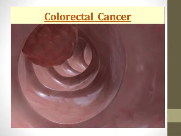
Pathology • More often sigmoid colon – logic of sigmoidoscopy • Most often single tumors, but multiple synchronous or metachronous tumors are not unusual
MACROSCOPY a)exofitic –cauliflower like b)schirous Major hyperplasie of fibroconjunctiv tissue Circular development – stenosis Fmore often left side c) coloide (mucinous): Proiferation of mucinous cells; Soft, friable, bleeding Often right side, young patients d) ulceration
Pathology • Microscopy: • adenocarcinoama: cylindric epithemlium • Carcinoid tumors – very unusual; • Epidermoid carcinoma– exceptional; • Sarcoama
a) direct: In the wall – serosa – ajacent organ In the surface: circumferential; longitudinal: Along submucosal layer b) lymphatic: Most often Intraperietal – local – regional lymph nodes c) Vascular: Colic veins – portal system – lver MTS Lombar and vertebral veins – pulmonary MTS d) intraluminal: Neoplastic cells get detached and reseaded (anastomotic recurrences) . e) transperitoneal: T4a – exposure to the serosa Douglas pouch Omentum Peritoneal carcinomkatosis f) perineural: . Spread pathways
Staging • Stadiul 0 Tis N0 M0 Dukes • Stadiul I T1 N0 M0 A T2 N0 M0 Stadiul II T3 N0 M0 B T4 N0 M0 Stadiul III any T N1 M0 C any T N2, N3 M0 • Stadiul IV any T any N M1
Changes in bowel habit Constipation Diarrhea !alternation of constipation with diarrhea) Dependent on location of the colon Pain From discomfort to colicky Aggressive peristalsis above the tumor Borborism Meteorism Can suggest the location Location of pain: RLQ – distension of cecum; Epigastric – often in transverse colon cancer; RUIQ lombar – may creat confusion Bleeding occult melenar; Hematochezia Other synptoms: ~ gastric problems ~ billiary symptoms ~ urinary syptoms General signs : anorexia, weight loss, low fever SYMPTOMS
Clinical examination • Often negative • GENERAL; • general: • Palor, apathy, diminshed turgor • Cachexia – advanced stages • LOCAL • Nothing • Tumor • Ascitis • Hepatomegaly • Rectal/vaginal : • Sigmoid tumors falling in the pouch of Douglas; • Carcinomatosis.
LAB • Non specific • Anaemia (microcytis, hypochromic) ; • Increased ESR • leucocitosis • Abnormal liver tests • CEA • Not for diagnostic purpose; • Only high values are significant for C colon, stomach, pancreas. Normal value do not have significance • More valuable for post therapy follow up • Occult blood test: screening ??? • Colic cytology
X-Ray • Corect dg in 90% • Plain X-Ray in complications • Barium enema • Wall rigidity • Filling defects. • Stenosis – golf trousers • Ulcerations
Colonoscopy • Biopsy • Treatment
Evolution and complications 1. Obstruction: • Left colon and rectum • Incomplete obstruction to acut obstruction • Typical presentation 2. Perforation: • a) extension through the wall; • b) diastatic: • c) juxtatumoral.
3. Septical: Abscess formation; Peritonitis 4. Fistula: exterior – piostercoral fistula Other organ; 5. Volvulus 6. Invagination 7. Compression 8. Invazia organelor învecinate: 9. Anemia: 10. Metastasis
TREATMENT • Surgical: - tumor, lymph nodes, regiomal lymphnodes +/- invadet organs • Radical – oncologic colectomy with regional
Paliative: • 1. by pass: • 2. diverting stoma • 3. stents