Download

1 / 59
600 likes | 920 Vues
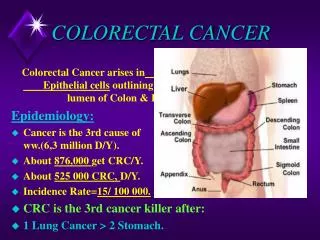
Colorectal Cancer. Brunner, pp. 1098-1107. Colorectal Cancer Statistics and Risk. Second leading cause of death from cancer Most are adenocarcinoma Approximately 70-75% occur in colon; 25-30% in rectum with ½ occurring in the rectosigmoid area

E N D
Colorectal Cancer Brunner, pp. 1098-1107
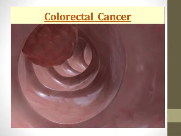
Colorectal Cancer Statistics and Risk • Second leading cause of death from cancer • Most are adenocarcinoma • Approximately 70-75% occur in colon; 25-30% in rectum with ½ occurring in the rectosigmoid area • Over ¾ of cancers come from polyps that spread into mucosal lining and into lymph system and then to liver*,lungs, bone, brain • Risk factors p. 1099, Chart 38-9
Manifestations • Maybe none for 5-15 years • Hematochezia or melena • Abdominal pain/cramping • Weakness, fatigue, anemia, weight loss • Change in bowel habits • Change in stool caliber • Fullness in lower abdomen or rectum or palpable mass
Complications • Intestinal obstruction (pain, vomiting, distention, unusual bowel sounds, no stool) • Iron-deficiency anemia from blood loss • Perforation with peritonitis (sudden pain, distention, fever, sepsis) • Fistula formation
Diagnostics • Colonoscopy is gold standard—polyps or tumors may be seen, but bx is confirmation. Starting at 50, then depending on findings, family hx—may be q 5 or q 10yr • Hemoccult or guaiac (FOB) • Barium enema • Labs: CBC, coag studies, liver functions, CEA—initial and to monitor treatment and recurrence • CT or MRI
Collaborative Care: Surgery • Treatment depends on Dukes or TNM classification • Polypectomy during colonoscopy for in-situ • Colon resection (right or left hemicolectomy) with end-to-end anastomosis with lymph removal (lap procedures decrease recovery time) • Abdominal-perineal resection with ostomy • A-P resection with temporary ostomy to preserve anal sphincter. May include construction of rectal pouch. • If metastasized, surgery may be palliative to control bleeding or obstructive sx
Chemo and Radiation Therapy • Treatment is highly individualized, but combo platters are usually used. Most common drugs: • 5-FU • Leucovorin • Xeloda • Mitomycin • Radiation as adjuvant or for metastasis to reduce tumor size & provide symptomatic relief
Nursing Management: History • Colon, breast, ovarian cancer, familial or hereditary polyposis, inflammatory bowel dz, meds affecting bowel function • High-fat, low-fiber diet • Weakness, fatigue, anorexia, wt loss, N/V • Bowel changes: urgency, bleeding, mucoid, black, gas, decrease in caliber, pain
Nursing Management: Objective Data • Pallor, cachexia, lymphadenopathy • Abd mass, distention, ascites, hepatomegaly • Hemoccult + stools, anemia • + DRE, + scopes, + radiography
Preop Nursing Management • Preop teaching—may need ostomy teaching by wound care or ostomy care nurse, preferably • Dietary modifications may be done several days before surgery • Need info about bowel prep procedure • Bowel cleansing and or antibiotics to decrease contamination • Maybe need TPN before surgery • Need a lot of emotional support
Postop Nursing Management • If reanastamosis is done, then postop care is routine abdominal surgery. Incision may be large, but closed with staples. Remember to check incision, dressing, and drainage. • Lap procedures will only have small midline incision and lap sites covered with Tegaderm • Pt may have NGT or TPN. May be NPO, ice chips, or clear liqs depending on type of surgery
Surgical Nsg Care cont’d • Monitor for infection in any skin break • Provide adequate pain control and give prophylacticly • Monitor for signals of readiness to resume oral intake • If abdominal-perineal surgery is done for extensive metastasis, care of both an abdominal and an open perineal wound and drain management is necessary. • Ostomy care if indicated • Probs with sexual dysfunction
Patient Education • For screening: • FOB q yr • Patients > 50 to have routine colonoscopy; 45 in blacks—repeat q 10 y unless + hx • Teaching regarding colonoscopy prep • Teach patients how to recognize early warning signs • For postop: • Home instruction on sitz baths, wound & ostomy care, dietary management • Don’t forget psychosocial issues, sexual concerns & grief mgmt
Prostate Cancer Brunner, pp. 1516-1530
Prostate Cancer • Most common cancer in men and • 2nd leading cause of death from cancer. 2/3 are over 65 y.o. • Almost 30,000 die each year. Interestingly, early dx leads to cure. • 5-year survival rate is 98%
Risk Factors • >50 y.o. • African American (twice as likely) • Family hx (father or brother twice as likely) • High fat diet, high red meat intake, Vitamin A supplements, low intake of fruits and vegs • Positive HPC1, BRCA1 and BRCA2 gene mutations
Manifestations of Prostate Cancer • Asymptomatic at 1st • Dysuria, urgency, frequency, hesitancy, dribbling, nocturia, retention, interrupted stream, inability to urinate, hematuria, oliguria • Painful ejaculation, back, hip, leg pain and weakness, and perineal or rectal discomfort • Anemia, nausea, wt loss
Complications • Metastasis to lymph nodes, bones, bladder, lungs, and liver • Bone mets are especially painful because of spinal cord compression and destruction of pelvic bone, femoral head, or lumbosacral spine. Pain control is important aspect of care.
Diagnostics • DRE reveals hard, nodular, asymmetrical gland • PSA>4 (not all elevations are cancer). For screening and monitoring success of tx • UA, CBC, Alkaline phosphatase • Transrectal US with needle bx • CT, MRI, bone scan
Medical Management of Prostate Cancer • Depends on stage • Pharmacologic: androgen deprivation therapy or androgen antagonist therapy. Accomplished by giving meds such as Lupron (testicular suppression of androgen), or Eulexin (adrenal suppression). • External beam or brachytherapy (internal radiation with seed implants)—with or without surgery • Cryotherapy—liquid nitrogen placed into prostate • Watchful waiting—more common in elderly
Surgical Management • Surgical tx includes radical prostatectomy (prostate, seminal vesicles, part of bladder neck and lymphs are removed) by one of three methods: suprapubic, retropubic, perineal—see p. 1525, Figure 49-4 • May also be done laproscopically and with nerve-sparing procedure • Orchiectomy may also be done if late stage (produces androgen suppression)
Complications • Urinary incontinence • Erectile dysfunction • Hemorrhage • Urinary retention • Infection • Dehiscence • DVT and PE
Nursing Management: Health Promotion • Teach importance of PSA and DRE beginning at age 50 and 45 for African Americans • If risk factors are present, screening may need to be done earlier • Teach symptoms of enlarged prostate and to seek help when it happens • Stress high success rate with early detection
Postop Nursing Management • Monitor for return of sensation from spinal anesthesia and protect from injury • Monitor 3-way Foley and CBI if used • Keep CBI running at rate that keeps urine pink without clots • Watch for hemorrhage • FF, keep strict I&O (subtract CBI) • Monitor surgical incision
Postop Nursing Care cont’d • After CBI is d/c, urine will be cranberry • Monitor for clots—call MD for irrigation order • Usually go home with cath; After cath is out, urine is racked (monitored by comparison samples) • Push fluids! Clots must be prevented • Expect bladder spasms and discomfort with first voiding which will be small • Give analgesics and also antispasmodics (if ordered), stool softeners • Emotional support
Patient/Family Education after Surgery • Catheter care and bag-switching • Kegel exercises • Wearing pad up to one year • Avoid intraabdominal pressure: Valsalva, lifting, long trips, strenuous activity, sitting or walking for long periods • Caffeine restriction, FF, urine will be cloudy • Watch for bright red bleeding, infection, decreased UOP, incision, calf tenderness • Management of ED—Viagra and penile implants
Breast Cancer Brunner, pp. 1481-1503
Overview * Factors used to help differentiate benign from malignant tumors include age, number of lumps, shape, consistency, mobility, tenderness, retraction. • BSE qmo beginning at age 20, but malignant lesions may not be palpable for 10 years; therefore mammography baseline 35-40 and qyr after 40. • Mutated cell doubles q30d; 30 doubling times for lump to get to 1 cm when it can become apparent
Breast Cancer Statistics • Most common 2nd to skin cancer • Highest death rate 2nd to lung cancer • Over 200,000 new cases; almost 41,000 deaths each year • Incidence is increasing; deaths decreasing especially among young women • Localized cancers without node involvement have 5-yr survival rate of 98%
Etiology and Risk Factors • Table 48-3 on p. 1483 shows gender, age, fa hx, personal hx, hormonal influences, parity, obesity, dietary factors, radiation exposure, and complicated benign disease as risk factors • Mutations in genes BRCA 1 and 2 increase risk, but can be reduced by having ovaries removed.
Protective Factors and Prevention Strategies • Full-term pregnancy before age 30 • Breastfeeding (delays exposure to estrogen) • Exercise after menopause • Close surveillance with high risk patients using MRI • Tamoxifen or Evista for high risk patients • Prophylactic mastectomy
Types of Breast Cancer • Ductal Carcinoma in Situ (noninvasive) • Confined to ducts • Mostly treated by simple mastectomy with radiation • Tamoxifen x 5 yrs for prophylaxis
Types cont’d • Invasive Carcinoma—Most serious: • Infiltrating ductal –80% of all breast cancers; very hard on palpation; more likely to metastasize to lung, bone, liver, brain; poorest prognosis. • Infiltrating lobular—10-15%; arise from thickened areas and may occur at several sites; may spread to above areas and meninges; poor prognosis.
Types cont’d • Invasive Carcinoma—Better outcomes: • Medullary—5%; encapsulated and large; fair prognosis. • Mucinous—3%; slow growing; good prognosis • Tubular—2%; metastasis rare; excellent prognosis
Types cont’d • Invasive Carcinoma—Rare, serious types: • Inflammatory—1-3%; causes pain, redness, enlarged and firm breast, edema, nipple retraction; attention is sought early; spreads quickly; chemo, radiation, surgery • Paget’s Disease—1%; ductal type; scaly lesion, burning, itching around nipple-areola area; bx is needed for dx; tx as above
Assessment (Chart 48-1, p. 1474) • Nontender • Fixed • Hard • Irregular border • Retraction • Dimpling • Usually upper outer quad • Lymphs, bone, lung sites most common sites of metastasis
Diagnostics • BSE: includes inspection & palpation • Mammography • US • MRI (for women at high risk) • Biopsy: definitive; can reveal type and stage and whether tumor is estrogen dependent
Breast Self-Exam (BSE), p. 1475-6 • Examine monthly , preferably after period, beginning at age 20 • Clinical exam q3yr 20-40; qyr after 40 • Examine in shower with soap and water • Look at breasts in mirror, then raise arms • Put hands on hips; then lean forward • Use a method to palpate entire breast tissue, including tail of Spence
Mammography • Detects tumors using x-ray even before they are palpable (usually 1 cm-10 years) • Can show early cancer tissue changes if compared to previous x-rays • Yearly mammography starting at 40 (talk with MD if high-risk)
Staging • Most women are Stage 2 @ time of dx • Survival Rates depend on: • Hormone receptors • Growth factor receptors (HER-2) • Tumor differentiation, size • Proliferation (number) • DNA content • Axillary node involvement
Other Management • Hormone suppression by oophrectomy, removal of pituitary gland, or adrenal glands • Radiation (internal and external)—only in breast conserving procedures or with chest wall involvement • Pharmacologic—Hormones if tumor is hormone dependent; Antineoplastics—3 of 5 drugs being used
Preop Nursing Management • Education about dx procedures, meds, postop wound care, managing chemo SEs, prosthetics • Physician will discuss treatment options and reconstruction • Emotional support—Use therapeutic communication and education to address many fears and anxieties r/t death, reoccurrence, txs, relationships, and finances
Postop Nursing Management • Pain management (paresthesia is common) • Meds • Arm elevation • Drain management • Management of incision and dressing • Arm exercises (1491) • Emotional support • Education for home management
Preventing Postop Complications • Hematoma—indicates internal hemorrhage. Monitor x 12h—if forms, call MD immediately • External hemorrhage • Infection—incision, etc. • Lymphedema—occurs more often in pts who have had axillary node dissection compared to sentinel node dissection • Injury and trauma to arm
Radiation • External beam—most common • Brachytherapy with implant into lumpectomy site • Intraoperative radiation therapy (IORT)—intense radiation to surgical site after lump is removed
Chemotherapy • Cytoxan, methotrexate, and fluorouracil regimen is most common • Taxol may be added for axillary node involvement • Hormonal therapy with Tamoxifen (estrogen blocker) for premenopausal women; Arimidex (enzyme inhibitor that prevents estrogen from forming) for postmenopausal • Targeted therapy using Herceptin which inactivates the HER-2 protein that makes tumor grow
Reconstruction • Enables women to maintain a sense of wholeness and to balance other breast • Some women prefer prosthetics • In most cases, can be done immediately or within one year of mastectomy • Done in stages • More successful if women have realistic expectations and have reconstruction done as soon as possible
Types of Reconstructive Surgery • Saline implant: • Temporary implant placed inside pectoralis muscle with port attached for injecting saline over a period of weeks. When tissue is stretched enough, permanent one is placed. • Advantages—office visits and OP surgery, less complications. • Disadvantages—less natural looking, synthetic material used