Download
1 / 24
260 likes | 1.12k Vues
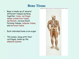
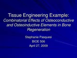
Tissue Engineering Example: Combinatorial Effects of Osteoconductive and Osteoinductive Elements in Bone Regeneration. Stephanie Pasquesi BIOE 506 April 27, 2009. Coating of VEGF-releasing scaffolds with bioactive glass for angiogenesis and bone regeneration.

E N D
Tissue Engineering Example:Combinatorial Effects of Osteoconductive and Osteoinductive Elements in Bone Regeneration Stephanie Pasquesi BIOE 506 April 27, 2009
Coating of VEGF-releasing scaffolds with bioactive glass for angiogenesis and bone regeneration J. Kent Leach a,b, Darnell Kaigler b, Zhuo Wang b, Paul H. Krebsbach b, David J. Mooney a,b a Division of Engineering and Applied Sciences, Harvard University, Cambridge, MA 02138, USA b School of Dentistry, Biologic and Materials Sciences, University of Michigan, Ann Arbor, MI 48109, USA Biomaterials (2006)
Definitions • Osteoconduction – the ability of some materials to serve as a scaffold to which bone cells can attach, migrate, grow, and divide • Osteoinduction – the capacity of normal chemicals in the body to stimulate primitive stem cells or immature bone cells to grow and mature, forming healthy bone tissue • Neovascularization – the formation of functional microvascular networks with red blood cell perfusion (i.e. formation of new blood vessels) • Different from angiogenesis: protrusion and outgrowth of capillary buds from pre-existing blood vessels • Mitogen – chemical substance, usually a protein, which promotes cell division and mitosis
Current Bone Graft Materials • Autografts • Graft tissue (bone) from the patient • Problems: chronic pain at site of bone harvestation, limited supply • Allografts • Graft tissue (bone) from someone other than the patient • Problems: immune rejection, risk of disease transmission • Metallic Implants • Problems: immune rejection, different mechanical properties, stress risers in existing bone, risk of poor positioning by surgeon, etc.
Synthetic Matrices • Scaffolds of synthetic material (effectively synthetic ECM) to provide a support for osteoblast proliferation in critically-sized bone defects • Once osteoblasts attach to scaffold and osteogenesis is induced, scaffold dissolves away, leaving new healed bone in its place • Currently can present osteoinductive growth factors from within matrices, but lack the osteoconductivity of conventional graft materials • Interest in developing a composite material allowing for delivery of osteoinductive macromolecules and possessing osteoconductive properties
VEGF • VEGF = vascular endothelial growth factor • Protein, endothelial cell mitogen • Well known for angiogenesis, also important in osteogenesis • Promotes neovascularization, bone turnover, osteoblast migration and mineralization • Osteoinductive
Bioactive Glass (BG) • Osteoconductive, surface active, glass-ceramic material composed of several oxidized minerals • Good adhesive bonding capacity with bone and some connective tissues • Some studies have shown it may exhibit both osteoconductive and osteoinductive properties • Hattar et al (2005), Bosetti et al (2005) • Others show stronger matrices and accelerated deposition of hydroxyapatite layer in vitro • Suggests improved integration upon placement in vivo • Maquet et al (2003), Verrier et al (2004), Lu et al (2005), Yao et al (2005)
Hypothesis • Adding an osteoconductive (BG) surface to VEGF (osteoinductive) releasing scaffolds serving as synthetic ECM will enhance bone regeneration through improved vascularization and integration with native tissues
Procedure Overview • VEGF incorporated into 3D porous scaffolds made from poly(lactide-co-glycolide) for localized protein delivery • Scaffold surface coated with bioactive glass to enhance osteoconductivity • Investigated in vitro models • HMVEC (human microvascular endothelial cell) proliferation • Progenitor cell differentiation • Investigated in vivo models • Neovascularization • Bone regeneration
Scaffold Fabrication • 3 μg VEGF incorporated in polymeric scaffolds by gas foaming/particulate leaching process • Scaffold coated with BG by soaking in ethanol to reduce hydrophobicity and then submerging in a BG slurry in distilled water • BG deposited was 0.5 +/- 0.2 mg BG particulate on scaffold.
Scaffold VEGF Release • VEGF released in a sustained fashion over 18 days • Radio-labeled VEGF was used as a tracer
In Vitro Testing • HMVECs grown in wells containing four different scaffold types • Uncoated Blank Scaffolds (BL) • Uncoated VEGF-releasing Scaffolds (V) • BG Coated Blank Scaffolds (BGBL) • BG Coated VEGF-releasing Scaffolds (BGV)
In Vitro Testing – HMVEC Proliferation • All groups compared to control demonstrated increased HMVEC proliferation through day 6 • Enhanced proliferation in BGBL samples was not detectable by day 9 • BG coating has an additive proliferation affect when comparing V to BGV samples • By days 10-12 proliferation rate of BGV decreased with respect to that of V Filled – BL (control) Open – V (VEGF, no coating) Horizontal Striped – BGBL (BG, no VEGF) Vertical Striped – BGV (BG and VEGF)
In Vitro Testing – Progenitor Cell Differentiation • Scaffolds were seeded with hMSCs (human mesenchymal stem cells) • Alkaline phosphatase expression • Indicator of progenitor cell differentiation • No significant differences between different scaffolds • Osteocalcin secretion • Secreted differentiation marker • No significant differences between different scaffolds Filled – BL, Open – V, Horizontal Striped – BGBL, Vertical Striped – BGV
In Vivo Testing • 9mm diameter hole made in Lewis rat crania • 2 types of implant • BG coated scaffold with VEGF (BGV) • BG coated control scaffold (BGC) • At 2 weeks, some rats euthanized and scaffolds scanned for neovascularization • At 12 weeks, other rats euthanized and scaffolds inspected for bone regeneration
In Vivo Testing - Neovascularization • 2 week samples were tested for the presence of blood vessels by immunostaining for vWF (von Willebrand Factor) • vWF: glycoprotein present in large quantities in subendothelial matrices • Vessels = circular, dark brown (arrows) BGC – top, BGV - bottom
In Vivo Testing - Neovascularization • Significantly more (p<0.001) vessels in BGV than BGC scaffolds • BGV displayed 117 ± 20 vessels/cm2 • BGC displayed 66 ± 8 vessels/cm2 • Area between dashed lines: scaffold alone • 36 ± 9 vessels/cm2 (unpublished)
In Vivo Testing – Bone Regeneration • 12 week samples were scanned for bone regeneration by microCT imaging • Left: Distribution of new mineralized tissue • Right: Nearly complete bridging of defect by new mineralized tissue
In Vivo Testing – Bone Regeneration • Bone volume fraction • BGV slightly higher, no significant difference • BGV: 20 ± 4% • BGC: 14 ± 6% • Bone Mineral Density • BGV shows significant increase (p=0.02) vs. BGC • BGV: 177 ± 17 mg/cm3 • BGC: 135 ± 27 mg/cm3 • Area between dashed lines: scaffold alone • 120 ± 20 mg/cm3(unpublished)
Conclusions - BG • BG coating induces significant increase in proliferation of endothelial cells in vitro and in vivo • Angiogenesis further increased with the delivery of VEGF from BG coated scaffolds • Large difference in masses of BG (500 μg) and VEGF (3 μg) needed for similar response • Suggests that angiogenic effects of BG may be indirect
Conclusions - BG • Did not show osteogenic response of BG unlike prior studies • Relatively low concentrations of BG used in this model were enough to elicit angiogenic response, higher concentrations may yield a more robust osteogenic reaction • Previous studies used larger concentrations, packing the defect area with BG • Osteoconductivity of BG was limited by dissolution rate of coating • BG coating offers inductive component not available through other osteoconductive materials
Conclusions - VEGF • Prolonged delivery of VEGF improves maturation of newly formed bone • Significant increase in bone mineral density • Slight increase in bone volume fraction • Expected from prior studies • Defect regeneration may benefit from localized VEGF presentation • Establishes a vascular network for nutrient transport, potentially supplying progenitor cells for healing
Overall Conclusions • Strong linkage between angiogenesis and bone regeneration • Combinatorial approaches of delivering osteoinductive factors from osteoconductive scaffolds provide therapeutic benefit • May achieve desired tissue response by capitalizing on degradation components of synthetic ECM and inductive factors released from the matrix