Download
1 / 70
700 likes | 716 Vues
Learn about the structure and function of arteries, capillaries, and veins, including the endothelium, lumen, connective tissue, and valves. Explore the role of vasoconstriction and vasodilation in controlling blood flow.

E N D
What you Should Know • The structure and function of arteries, capillaries and veins including the endothelium, central lumen, connective tissue, elastic fibres, smooth muscle and valves. • The role of vasoconstriction and vasodilation in controlling blood flow.
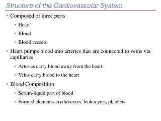
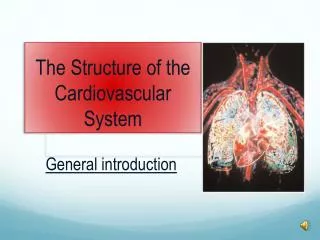
What is the Cardiovascular system? • The cardiovascular system, also known as the circulatory system, is composed of blood, blood vessels and the heart. • The heart functions as a pump to move blood through the blood vessels of the body. • A circulatory system is essential for large, multi-cellular organisms, such as humans and animals, and provide at least five major functions that are necessary for life.
The five major functions of the cardiovascular system are: • Transporting oxygen and removing carbon dioxide • Transporting nutrients and removing wastes • Fighting disease • Transporting hormones • Regulating body temperature
Cardiovascular system • http://www.youtube.com/watch?v=CjNKbL_-cwA
Components of the CVS • The CVS consists of a double pump (the heart) and a complex system of blood vessels.
Blood circulates from the heart through the arteries to the capillaries to the veins and back to the heart. • There is a decrease in blood pressure as the blood moves away from the heart.
Blood Vessels • There are 3 types of blood vessel: • Arteries (and arterioles) • Capillaries • Veins (and venules)
Central lumen of blood vessels • The endothelium lining the central lumen of the blood vessels is surrounded by layers of tissue. • These surroundings layer differ in each type of blood vessel
ARTERIES Carry blood away from the heart Endothelium One cell thick Elastic tissues & smooth muscles Rebounds Evens flow Fibrous tissue Tough Resists stretch
Arteries • Arteries carry blood away from the heart. • The largest arteries e.g. the Aorta, have thick elastic walls which can stretch to accommodate the surge of blood after each contraction of the heart. • Arteries branch many times, forming smaller and smaller vessels, the smallest of which are arterioles. • Contraction of the smooth muscle lining the walls of the arterioles allows them to open or close to varying degrees to adjust blood flow to different parts of the body e.g. during vasodilation and vasoconstriction
VEINS Carry blood towards the heart Endothelium Larger lumen than arteries Thinner muscle layer & few elastic fibres Blood at lower pressure Fibrous tissue
VEINS Contain valves Prevents backflow of blood Situated between skeletal muscles Muscle compresses vein when contracted Blood “squirted” towards heart
CAPILLARIES Transport blood between arteries and veins Form large networks (capillary beds) Exchange of materials between blood and cells Their walls are only one cell thick, allowing nutrients and waste to diffuse through with ease.
Capillaries Arteriole Venule Capillaries (capillary bed)
What you Should Know • Tissue fluid supplies cells with glucose, oxygen and other substances. • Carbon dioxide and other metabolic wastes diffuse out of the cells and into the tissue fluid to be excreted. • Much of the tissue fluid returns to the blood. • Lymphatic vessels absorb excess tissue fluid and return the lymph fluid to the circulatory system. • Similarity of tissue fluid and blood plasma • Pressure filtration of fluids through capillary walls.
Tissue Fluid • Blood consists of red and white blood cells, platelets and plasma • Plasma is a watery yellow liquid containing dissolved substances such as glucose, amino acids, respiratory gases, plasma proteins and useful ions
The blood plasma fluid which leaks into the tissues is called tissue fluid • Some of the tissue fluid returns to the blood at the venous end of the capillary
Cells tissue organs Lymphatic system
Blood plasma to tissue fluid • Blood pressure forces the fluid part of the blood along with small soluble molecules out of the capillaries into the tissue fluid. • Blood cells and large protein molecules are left behind • The cells exchange molecules with the tissue fluid by diffusion down a concentration gradient • Useful molecules such as food and oxygen diffuse into the cells whilst carbon dioxide and wastes diffuse out of them
Lymph • The fluid that does not return to the blood is now referred to as lymph and is collected by the lymphatic system. • Lymph travels under low pressure to enter the main circulation near the heart • The lymphatic system has no pump • The contraction of skeletal muscles squeezes lymph along the vessels • Lymph travels through vessels in one direction only due to the presence of valves
Summary Tissue Fluid and Lymphatic System Lymph passes into lymphatic system Blood arriving in the arteriole high pressure Lymph vessel Blood leaving in venule low pressure Some tissue fluid enters capillary by osmosis capillary Some tissue fluid enters lymphatic system Some plasma forced out of capillary Respiring cell Tissue fluid
What You Should Know • Definition of cardiac output and its calculation. • Description of the cardiac cycle to include the functions of atrial systole, ventricular systole, diastole. • Effect of pressure changes on atrio-ventricular (AV) and semi lunar (SL) valves.
Heart Rate (HR) • Number of times heart beats in one minute • Normal values around 72bpm • Normal range is between 60-90
Cardiac Output Cardiac Output is the volume of blood pumped by each ventricle per minute and is the function of two factors: • Heart rate (beats per minute) • Stroke volume (the volume of blood ejected by each ventricle during each contraction) • The left and right ventricles pump the same volume of blood through the aorta and pulmonary artery. CO = HR x SV
Cardiac Output (CO) • The volume of blood pumped by each ventricle per minute, measured in litres • Calculated as follows • CO = HR x SV • Normal values are around 5 litres/min
Stroke Volume (SV) • Volume of blood ejected by each ventricle during contraction • Normal values are around 70ml
At rest: HR = 72bpm SV = 70ml i.e. CO = 72 x 70 = 5040 ml/min = 5 litres/min • Cardiac Output varies between individuals and depends on their physical fitness and level of activity. • For example, the heart of a highly trained athlete can pump 30-35 l/min • Most non-athletes can only achieve a max of 20 litres.
Some typical values for cardiac output at varying levels of activity
Revision on Circulatory System Double system The pulmonary circuit carries deoxygenated blood from the right ventricle to the lungs and returns oxygenated blood to heart The systemic circuit carries oxygenated blood from the left ventricle to the aorta and then the rest of the body and returns deoxygenated from the body to the heart
The cardiac cycle Each heartbeat is called a cardiac cycle and consists of the following Atria contract simultaneously Ventricles contract simultaneously All chambers relax Lasts about 0.8 secs (0.3systole,0.5 diastole)
Two phases of the cardiac cycle Systole: contraction of the heart (Atrial first, the ventricular), blood forced out of chambers Diastole: relaxation of the heart, chambers fill with blood
The opening and closing of the AV and SL valves are responsible for the heart sounds heard with a stethoscope.
THE CARDIAC CYCLE • Atrial & ventricular diastole • Atrial systole, ventricular diastole • Ventricular systole, atrial diastole
ATRIAL & VENTRICULAR DIASTOLE • Blood enters atria from vena cava & pulmonary vein • AV valves open
ATRIAL SYSTOLE & VENTRICULAR DIASTOLE • Both atria contract • Blood forced into relaxed ventricles • AV valves still open • Ring of muscle around entrance to each atrium closed • Prevents backflow of blood into veins
ATRIAL DIASTOLE & VENTRICULAR SYSTOLE • About 0.1 secs after atrial systole • Ventricles contract • Blood forced into arteries through the open semi-lunar valves • AV valves close • Prevents backflow of blood to atria
ATRIAL DIASTOLE & VENTRICULAR SYSTOLE • Semi-lunar valves close when pressure in arteries exceeds pressure in ventricles • Cardiac cycle begins again!