Download

1 / 62
710 likes | 1.76k Vues
Structure and Function of the Muscular, Neuromuscular, Cardiovascular, and Respiratory Systems. chapter 1. Structure and Function of the Muscular, Neuromuscular, Cardiovascular, and Respiratory Systems. Gary R. Hunter, PhD, CSCS, FACSM Robert T. Harris, PhD. Chapter Objectives .

E N D
Structure and Function of the Muscular, Neuromuscular, Cardiovascular, and Respiratory Systems chapter1 Structure and Function of the Muscular, Neuromuscular, Cardiovascular, and Respiratory Systems Gary R. Hunter, PhD, CSCS, FACSMRobert T. Harris, PhD
Chapter Objectives • Describe the macrostructure and micro-structure of muscle. • Describe the sliding-filament theory. • Describe the characteristics of different muscle fiber types. • Describe the characteristics of the cardio-vascular and respiratory systems.
Section Outline • Muscular System • Macrostructure and Microstructure • Sliding-Filament Theory of Muscular Contraction • Resting Phase • Excitation-Contraction Coupling Phase • Contraction Phase • Recharge Phase • Relaxation Phase
Muscular System • Macrostructure and Microstructure • Each skeletal muscle is an organ that contains muscle tissue, connective tissue, nerves, and blood vessels. • Fibrous connective tissue, or epimysium, covers the body's more than 430 skeletal muscles.
Schematic Drawing of a Muscle • Figure 1.1 (next slide) • Schematic drawing of a muscle illustrating three types of connective tissue: • Epimysium (the outer layer) • Perimysium (surrounding each fasciculus, or group of fibers) • Endomysium (surrounding individual fibers)
Motor Unit • Figure 1.2 (next slide) • A motor unit consists of a motor neuron and the muscle fibers it innervates. • There are typically several hundred muscle fibers in a single motor unit.
Muscle Fiber • Figure 1.3 (next slide) • Sectional view of a muscle fiber
Myosin and Actin • Figure 1.4 (next slide) • The slide shows a detailed view of the myosin and actin protein filaments in muscle. • The arrangement of myosin (thick) and actin (thin) filaments gives skeletal muscle its striated appearance.
Key Point • The discharge of an action potential from a motor nerve signals the release of calcium from the sarcoplasmic reticulum into the myofibril, causing tension development in muscle.
Muscular System • Sliding-Filament Theory of Muscular Contraction • The sliding-filament theory states that the actin filaments at each end of the sarcomere slide inward on myosin filaments, pulling the Z-lines toward the center of the sarcomere and thus shortening the muscle fiber.
Contraction of a Myofibril • Figure 1.5 (next slide) • (a) In stretched muscle the I-bands and H-zone are elongated, and there is low force potential due to reduced cross-bridge–actin alignment. • (b) When muscle contracts (here partially), the I-bands and H-zone are shortened. • (c) With completely contracted muscle, there is low force potential due to reduced cross-bridge–actin alignment.
Muscular System • Sliding-Filament Theory of Muscular Contraction • Resting Phase • Excitation-Contraction Coupling Phase • Contraction Phase • Recharge Phase • Relaxation Phase
Section Outline • Neuromuscular System • Activation of Muscles • Muscle Fiber Types • Motor Unit Recruitment Patterns During Exercise • Preloading • Proprioception • Muscle Spindles • Golgi Tendon Organs • Older Muscle
Neuromuscular System • Activation of Muscles • Arrival of the action potential at the nerve terminal causes the release of acetylcholine. Once a sufficient amount of acetylcholine is released, an action potential is generated across the sarco-lemma, and the fiber contracts. • The extent of control of a muscle depends on the number of muscle fibers within each motor unit. • Muscles that function with great precision may have as few as one muscle fiber per motor neuron. • Muscles that require less precision may have several hundred fibers served by one motor neuron.
Key Term • all-or-none principle: All of the muscle fibers in the motor unit contract and develop force at the same time. There is no such thing as a motor neuron stimulus that causes only some of the fibers to contract. Similarly, a stronger action potential cannot produce a stronger contraction.
Stimulated Motor Unit • Figure 1.6 (next slide) • Twitch, twitch summation, and tetanus of a motor unit: • a = single twitch • b = force resulting from summation of two twitches • c = unfused tetanus • d = fused tetanus
Neuromuscular System • Muscle Fiber Types • Type I (slow-twitch) • Type IIa (fast-twitch) • Type IIab (fast-twitch); now named as Type IIax • Type IIb (fast-twitch); now named as Type IIx
Key Point • Motor units are composed of muscle fibers with specific morphological and physio-logical characteristics that determine their functional capacity.
Neuromuscular System • Motor Unit Recruitment Patterns During Exercise • The force output of a muscle can be varied through change in the frequency of activation of individual motor units or change in the number of activated motor units.
Neuromuscular System • Preloading • Occurs when a load is lifted, since sufficient force must be developed to overcome the inertia of the load • Proprioception • Information concerning kinesthetic sense, or conscious appreciation of the position of body parts with respect to gravity • Processed at subconscious levels
Key Point • Proprioceptors are specialized sensory receptors that provide the central nervous system with information needed to maintain muscle tone and perform complex coordi-nated movements.
Neuromuscular System • How Can Athletes Improve Force Production? • Recruit large muscles or muscle groups during an activity. • Increase the cross-sectional area of muscles involved in the desired activity. • Preload a muscle just before a concentric action to enhance force production during the subsequent muscle action. • Use preloading during training to develop strength early in the range of motion.
Neuromuscular System • Proprioception • Muscle Spindles • Muscle spindles are proprioceptors that consist of several modified muscle fibers enclosed in a sheath of connective tissue.
Muscle Spindle • Figure 1.7 (next slide) • When a muscle is stretched, deformation of the muscle spindle activates the sensory neuron, which sends an impulse to the spinal cord, where it synapses with a motor neuron, causing the muscleto contract.
Neuromuscular System • Proprioception • Golgi Tendon Organs (GTO) • Golgi tendon organs are proprioceptors located in tendons near the myotendinous junction. • They occur in series (i.e., attached end to end) with extrafusal muscle fibers.
Golgi Tendon Organ • Figure 1.8 (next slide) • When an extremely heavy load is placed on the muscle, discharge of the GTO occurs. • The sensory neuron of the GTO activates an inhibitory interneuron in the spinal cord, which in turn synapses with and inhibits a motor neuron serving the same muscle.
Neuromuscular System • Older Muscle • Muscle function is reduced in older adults. • Reductions in muscle size and strength are amplified in weight-bearing extensor muscles. • Muscle atrophy with aging results from losses in both number and size of muscle fibers, especially Type II muscle fibers. • Inactivity plays a major role but cannot account for all of the age-related loss of muscle and function.
Section Outline • Cardiovascular System • Heart • Valves • Conduction System • Electrocardiogram • Blood Vessels • Arteries • Capillaries • Veins • Blood
Cardiovascular System • Heart • The heart is a muscular organ made up of two interconnected but separate pumps. • The right ventricle pumps blood to the lungs. • The left ventricle pumps blood to the rest of the body.
Heart and Blood Flow • Figure 1.9 (next slide) • Structure of the human heart and course of blood flow through its chambers
Cardiovascular System • Heart • Valves • Tricuspid valve and mitral (bicuspid) valve • Aortic valve and pulmonary valve • Valves open and close passively, depending on the pressure gradient • Conduction System • Controls the mechanical contraction of the heart
Electrical Conduction System • Figure 1.10 (next slide) • The electrical conduction system of the heart
Cardiac Impulse • Figure 1.11 (next slide) • Transmission of the cardiac impulse through the heart, showing the time of appearance (in fractionsof a second) of the impulse in different parts of the heart
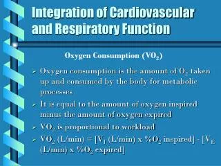
Cardiovascular System • Heart • Electrocardiogram • Recorded at the surface of the body • A graphic representation of the electrical activity of the heart
Electrocardiogram • Figure 1.12 (next slide) • Normal electrocardiogram
Cardiovascular System • Blood Vessels • Blood vessels operate in a closed-circuit system. • The arterial system carries blood away from the heart. • The venous system returns blood toward the heart.