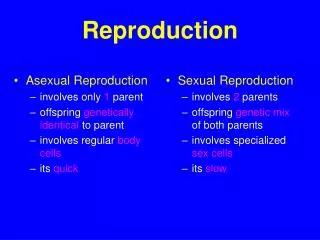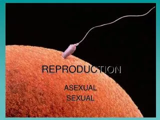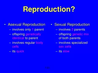
Reproduction
Reproduction. Female http://trc.ucdavis.edu/mjguinan/apc100/modules/Reproductive/mammal/female.html. The reproductive system of female mammals consists of the: ovaries, oviducts, uterus, vagina, and external genitalia (vulva, etc). Overview.

Reproduction
E N D
Presentation Transcript
Reproduction Female http://trc.ucdavis.edu/mjguinan/apc100/modules/Reproductive/mammal/female.html
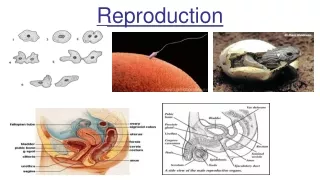
The reproductive system of female mammals consists of the: • ovaries, • oviducts, • uterus, • vagina, and • external genitalia (vulva, etc).
Overview • The compact ovary contains thousands of ovarian follicles, of which a few at a time rapidly go through several stages of maturation before ovulation. • The ova is transported to the uterus via the oviduct where, if fertilized, it implants, forms a placenta, develops, and is delivered via the vagina.
Overview-continued • The entire duct system is lined by epithelia with regional specializations. • There are a variety of species differences in the shape of the uterus, and the type of placentation that supports the embryo. • In contrast to the male, the urinary tract uses separate ducts until reaching the vestibule of the vagina.
Mammal, female The mammalian female reproductive tract consists of paired ovaries and a multi-part duct system.
Vestibule The vestibule is the terminal part of the duct system. It is shared with the urinary system
Vagina The vagina is a thick-walled fibromuscular tube that extends from the cervix to the vulva. The walls consist of a mucosa, muscularis, and adventitia.
The urethra from the urinary bladder joins with the reproductive duct in female mammals.
Cervix The cervix is a muscular valve which separates the vagina from the uterus. It may produce a mucus plug during infertile periods.
Uterus - body The body of the uterus is the site of implantation and development in primates.
Uterine horns Uterine horns provide additional space for the young to implant and develop. The length of the uterine horns varies between species, and may be nearly absent in primates.
Oviduct (fallopian tube) The uterine (fallopian) tube carries the ova to the uterus.
Infundibulum The infundibulum is the initial part of the duct system
Ovaries The ovaries are the female gonads. They produce the ova.
Ovaries • Produce 1 ova per cycle in cow • Produces 12-20 ova per cycle in sow • Produces 1-3 ova per cycle in ewe
The bovine uterus has two horns. • Both ovaries are shown and the one on your left has a corpus luteum present. • Developing follicles may be seen as little bumps on the surface of the ovary on the right.
Uterus The uterus, with layers called endometrium, myometrium and perimetrium, respectively.
Cervix CERVIX. The cervix acts as the valve of the reproductive tract. It separates the vagina from the uterus and is usually closed except during estrus and parturition.
Ovaries • OVARY/FOLLICLE. The two ovaries in mammals are the site of oogenesis, production of the female gametes. • In most mammals the OOGONIA have formed primordial follicles at the time of birth. These primordial follicles will go through several developmental stages until a mature follicle ruptures, expelling the ovum. • The ovaries also produce hormones important to reproductive cyclesand pregnancy.
The PRIMARY FOLLICLE develops from the primordial follicle and is characterized by several layers of follicular cells surrounding the oocyte. The follicular cells become more cuboidal and secretory in appearance and are referred to as granulosa cells. At this time, a clear layer of extracellular material, called the zona pellucida, can be seen around the oocyte.
PRIMORDIAL FOLLICLE. In most mammals the OOGONIA have formed primordial follicles at the time of birth. It is generally believed that all of the primordial follicles that are going to develop are present at the birth of the animal. The primordial follicle is a single egg or oocyte surrounded by a single layer of flattened cells called the follicular cells. Many of the follicles will not develop and will undergo a gradual breakdown or atrophy: this process is called atresia and the follicles are said to be atretic.
SECONDARY FOLLICLES develop from primary follicles and are characterized by the formation of an antrum. • The antrum is a fluid-filled space which begins as a number of small spaces between the granulosa cells. • As the several spaces unite into a single fluid-filled chamber, the oocyte remains on a mound of granulosa cells, the cumulus oophorus, to one side of the follicle. • A layer of granulosa cells remains surrounding the oocyte. • The cells secrete the female sex steroids.
Regressing CL’s • The corpus luteum forms from the cells of the theca interna and the granulosa cells under the influence of the pituitary luteinizing hormone.
Blood vessels and connective tissue grow into the luteal cells to form an endocrine gland which secretes progestins. • Hormones from the corpus luteum will support fetal development during pregnancy. • Afterwards it will become a scar (corpus albicans).
Estrous Cycle • Begins with puberty • Prepares the female for pregnancy • 4 periods to cycle • Proestrus • Estrus • Metestrus • Diestrus
Proestrus • follicular development from FSH • Estrogen increasing
Estrus • Female sexually receptive (standing heat) • estrogen levels high • length ~ 12 hrs to several days • surge of LH & FSH • Ovulation late or following estrus
Metestrus • CL (corpus luteum) forms • Produces progesterone
Diestrus • If pregnanacy occurs, CL remains • progesterone high • if no pregnancy, prostaglandin from uterus causes CL regression
Species cycle heat time of time to d hr ovulation mate------------------------------------------------------------------------- Cow 21 12-18 12-15 hr post-E 4-8 after E END Sow 20-21 48-72 18-40 h after 1st d & again 2nd start of estrus Ewe 16-17 24-36 18-26 h after start 12-18 after start Goat 19-20 34-39 9-19 h after start alternate days Mare 19-23 90-170 1d before to 1 d alternate days after estrus
Development of Follicle from FSH Ovulation, from LH surge CL Develops progesterone keeps animal preg. CL regresses to corpus albicans cycle begins again ANESTRUS period of no cycling Polyestrous vs seasonally polyestrous cow vs sheep Estrous Cycle Review
GnRH hypothalamus controls LH & FSH FSH stimulates follicle LH triggers ovulation & CL Estrogen from follicle, prepares for preg. Progesterone from CL prevents further cycling maintains pregnancy Prostaglandin (Pf2) regresses CL HORMONES - review
PREGNANCY • Length of Pregnancy, days • cow 282, 9 mo, 12 days • sow 114, 3 mo, 3 wk, 3 days • ewe 150, 5 mo • mare 336, 11 mo +
Sperm combines with ovum • ONLY 1 sperm can enter ovum • Cell divisions start • sometimes split, forming identical twins • Fertilization takes place in oviduct • Fertilized ovum moves down to uterine horn (or uterus) for implantation
Placentation • After implantation into the uterine endometrium, the developing embryo becomes surrounded by the chorionic, amniotic, and allantoic membranes. • In fish and birds these same membranes allow diffusive exchange of of CO2 and O2 with the environment, while nutrients are provided by the yolk sac.
In mammals, nutrients and wastes are exchanged with the maternal circulation across a specialized region of these these membranes called the placenta. • The placenta provides a large surface area of close association between the two independant circulations for diffusive and carrier mediated exchange.
In addition the placenta produces hormones to support the pregnancy. • The gross arrangement of the placenta may be diffuse, zonary discoid, or cotyledonary, depending on the species.
Bovine placenta Allantoic sac Amniotic sac
Fetal cotyledons are seen on the surface of the chorioallantoic membrane which attach to the maternal caruncles (maternal cotyledons) to provide for gas and nutrient exchange. The fetal cotyledon is shown here with villi, which develop off the surface of the chorio-allantoic membrane and grow into sulci of the maternal caruncle.This interdigitation is the site of blood gas and nutrient exchange. (There is no mixing of blood between the fetus and the mother.)
Parturition (giving birth) • Placenta produces more estrogen • Causes prostaglandin from uterine wall • Regression of CL, reduced progesterone • RELAXIN causes pelvic muscles, ligaments to ‘loosen’ • PROLACTIN stimulates milk synthesis • Oxytocin stimulates & strenghtens uterine contractions
Orientation at birth rear first one leg back head twisted too large inadequate contraction strength too long a birth process stillborn Membranes cover nose, prevent breathing Retained placenta Abnormal bleeding Premature births Difficulties at Birth
Female Reproduction Review • The ovaries contain a large number of follicles, of which a few rapidly mature during each cycle. The theca and granulosa cells proliferate as each follicle develops from a primordial, to primary, to secondary, to mature follicle (Graafian) which releases the oocyte and then transforms into a corpus luteum and later a corpus albicans.
The ova move into the first part of the uterine tube (oviduct, fallopian tube) where fertilization may take place. Implantation and embryo development occurs in the uterus which has a nourishing, glandular epithelia (endometrium) surrounded by smooth muscle (myometrium). • The uterus of some species have elongated horns or a dividing septum.