Cell Theory and Microscopes
Intro to the light Microscope Homework. INTRODUCTION TO THE LIGHT MICROSCOPE --Internet LabFamiliarize yourself with the microscope, run the tutorial and examine the parts you will be working with.Complete handout activitieshttp://www.udel.edu/biology/ketcham/microscope/Due Date: ____________.

Cell Theory and Microscopes
E N D
Presentation Transcript
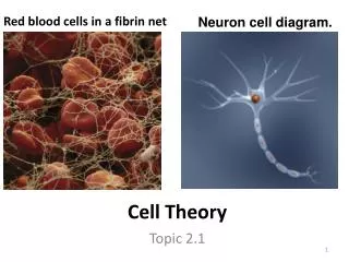
1. Cell Theory and Microscopes 41. Explain with evidence how the theories about life changed over time.
42. State the cell theory and explain how the invention of the microscope helped the development of the theory
S.C.H.1.4.1
S.C.H.1.4.2
S.C.H.1.4.3
S.C.H.3.4.2
B. Thomas
2. Intro to the light Microscope Homework INTRODUCTION TO THE LIGHT MICROSCOPE --Internet Lab�
Familiarize yourself with the microscope, run the tutorial and examine the parts you will be working with.
Complete handout activities
http://www.udel.edu/biology/ketcham/microscope/
Due Date: ____________
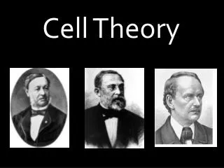
3. History of Cell Theory - Janssen Credit for the first compound (more than one lens) microscope is usually given to Zacharias Jansen, of Middleburg, Holland, around the year 1595.
Janssen and his father made a compound microscope by placing two convex lenses at each end of a tube Credit for the first compound (more than one lens) microscope is usually given to Zacharias Jansen, of Middleburg, Holland, around the year 1595.
Since Jansen was very young at that time, it's possible that his father Hans made the first one, but young Jansen perfected the production.
Details about the first Jansen microscopes are not clear, but there is some evidence which allows us to make some guesses about them Credit for the first compound (more than one lens) microscope is usually given to Zacharias Jansen, of Middleburg, Holland, around the year 1595.
Since Jansen was very young at that time, it's possible that his father Hans made the first one, but young Jansen perfected the production.
Details about the first Jansen microscopes are not clear, but there is some evidence which allows us to make some guesses about them
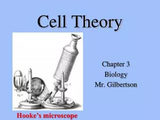
4. History of Cell Theory - Hooke In 1663 an English scientist, Robert Hooke, discovered cells in a piece of cork, which he examined under his primitive microscope
Hooke observed small empty like structures which he named cells
The cells that Hooke observed were non-living Hooke drew the cells he saw and also coined the word CELL. The word cell is derived from the Latin word 'cellula' which means small compartment.Hooke drew the cells he saw and also coined the word CELL. The word cell is derived from the Latin word 'cellula' which means small compartment.
5. History of Cell Theory - van Leeuwenhoek Anton van Leeuwenhoek was the first to observe and describe magnified living things with simple microscope. (working between 1670s-1680s) Leeuwenhoek was the first to see bacteria from teeth scrapings and animal-like protist from pond water Hooke used his own (single lens) monocular microscopes and was the first person to observe bacteria and protozoa. Leeuwenhoek is known to have made over 500 "microscopes," of which fewer than ten have survived to the present day. Leeuwenhoek's skill at grinding lenses, together with his naturally acute eyesight and great care in adjusting the lighting where he worked, enabled him to build microscopes that magnified over 200 times, with clearer and brighter images than any of his colleagues at that time. Leeuwenhoek looked at animal and plant tissues, at mineral crystals, and at fossils. He was the first to see microscopic single celled protists with shells, the foraminifera, which he described as "little cockles. . . no bigger than a coarse sand-grain." He discovered blood cells, and was the first to see living sperm cells of animals. He discovered microscopic animals such as nematodes (round worms) and rotifers.
Between 1680 and the early 1800's it appears that not much was accomplished in the study of cell structure. This may be due to the lack of quality lens for microscopes and the dedication to spend long hours of detailed observation over what microscopes existed at that time. Leeuwenhoek did not record his methodology for grinding quality lenses and thus microscopy suffered for over 100 years.
http://fig.cox.miami.edu/~cmallery/150/unity/cell.text.htm#text1Hooke used his own (single lens) monocular microscopes and was the first person to observe bacteria and protozoa. Leeuwenhoek is known to have made over 500 "microscopes," of which fewer than ten have survived to the present day. Leeuwenhoek's skill at grinding lenses, together with his naturally acute eyesight and great care in adjusting the lighting where he worked, enabled him to build microscopes that magnified over 200 times, with clearer and brighter images than any of his colleagues at that time. Leeuwenhoek looked at animal and plant tissues, at mineral crystals, and at fossils. He was the first to see microscopic single celled protists with shells, the foraminifera, which he described as "little cockles. . . no bigger than a coarse sand-grain." He discovered blood cells, and was the first to see living sperm cells of animals. He discovered microscopic animals such as nematodes (round worms) and rotifers.
Between 1680 and the early 1800's it appears that not much was accomplished in the study of cell structure. This may be due to the lack of quality lens for microscopes and the dedication to spend long hours of detailed observation over what microscopes existed at that time. Leeuwenhoek did not record his methodology for grinding quality lenses and thus microscopy suffered for over 100 years.
http://fig.cox.miami.edu/~cmallery/150/unity/cell.text.htm#text1
6. History of Cell Theory - Brown Around 1833 Robert Brown reported the discovery of the nucleus.
In the course of his microscopic studies of the epidermis of orchids, discovered in these cells "an opaque spot," which he named the nucleus Doubtless the same "spot" had been seen often enough before by other observers, but Brown was the first to recognize it as a component part of the vegetable cell and to give it a name.
LEFT) Robert Brown's Microscope
This is the instrument with which Robert Brown studied Brownian Movement and which he used in his work on identifying the nucleus of the living cell. This instrument is preserved at the Linnean Society in London. It is made of brass and is mounted onto the lid of the box in which it can be stored. For many years it was believed that Brown could not have seen the nucleus with such a primitive instrument.
�(RIGHT) Orchid cells under Brown's microscope
This is the view Brown obtained in 1828, when he first recognized the cell nucleus. It shows about twenty orchid epidermal cells, and the nucleus can clearly be seen within each cell. Three stomata can also be clearly seen - these are the breathing pores through which a plant exchanges gases with the atmosphere.�Doubtless the same "spot" had been seen often enough before by other observers, but Brown was the first to recognize it as a component part of the vegetable cell and to give it a name.
LEFT) Robert Brown's Microscope
This is the instrument with which Robert Brown studied Brownian Movement and which he used in his work on identifying the nucleus of the living cell. This instrument is preserved at the Linnean Society in London. It is made of brass and is mounted onto the lid of the box in which it can be stored. For many years it was believed that Brown could not have seen the nucleus with such a primitive instrument.
�(RIGHT) Orchid cells under Brown's microscope
This is the view Brown obtained in 1828, when he first recognized the cell nucleus. It shows about twenty orchid epidermal cells, and the nucleus can clearly be seen within each cell. Three stomata can also be clearly seen - these are the breathing pores through which a plant exchanges gases with the atmosphere.�
7. History of Cell Theory - Schleiden It was the German professor of botany at the University of Jena, Dr. M. J. Schleiden, who brought the nucleus to popular attention, and to asserted its all-importance in the function of a cell.(1838)
He came to believe that the nucleus is really the most important portion of the cell, in that it is the original structure from which the remainder of the cell is developed chleiden freely acknowledged his indebtedness to Brown for first knowledge of the nucleus, but he soon carried out his own observations of the nucleus, far beyond those of Brownchleiden freely acknowledged his indebtedness to Brown for first knowledge of the nucleus, but he soon carried out his own observations of the nucleus, far beyond those of Brown
8. History of Cell Theory - Schwann The following year, Dr Theodor Schwann (1839) who worked with animals, stated that all animals are made of cells.
Schwann published a book on animal and plant cells. He summarized his observations into three conclusions about cells:
1) The cell is the unit of structure, physiology, and organization in living things.
The cell retains a dual existence as a distinct entity and a building block in the construction of organisms.
Cells form by free-cell formation, similar to the formation of crystals (spontaneous generation).
Question: Which of these three conclusions do we know not to be true? Schwann was puzzling over certain details of animal histology which he could not clearly explain. He had noted a strange resemblance of embryonic cord material, from which the spinal column develops, to vegetable cells. Schwann recognized a cell-like character of certain animal tissues. Schwann felt that this similarity could not be mere coincidence, and it seemed to fit when Schleiden called his attention to the nucleus. Then at once he reasoned that if there really is the correspondence between vegetable and animal tissues that he suspected, and if the nucleus is so important in the vegetable cell as Schleiden believed, the nucleus should also be found in the ultimate particles of animal tissues. A closer study of animal tissues under the microscope showed, in particular in embryonic tissues, that the "opaque spots" that Schleiden described were found in abundance. The location of these nuclei at comparatively regular intervals suggested that they are found in definite compartments of the tissue, as Schleiden had shown to be the case with vegetables; indeed, the walls that separated such cell-like compartments one from another were in some cases visible. Soon Schwann was convinced that his original premise was right, and that all animal tissues are composed of cells not unlike the cells of vegetables. Adopting the same designation, Schwann propounded what soon became famous as the CELL THEORY. So expeditious was his observations that he published a book early in 1839, only a few months after the appearance of Schleiden's paperSchwann was puzzling over certain details of animal histology which he could not clearly explain. He had noted a strange resemblance of embryonic cord material, from which the spinal column develops, to vegetable cells. Schwann recognized a cell-like character of certain animal tissues. Schwann felt that this similarity could not be mere coincidence, and it seemed to fit when Schleiden called his attention to the nucleus. Then at once he reasoned that if there really is the correspondence between vegetable and animal tissues that he suspected, and if the nucleus is so important in the vegetable cell as Schleiden believed, the nucleus should also be found in the ultimate particles of animal tissues. A closer study of animal tissues under the microscope showed, in particular in embryonic tissues, that the "opaque spots" that Schleiden described were found in abundance. The location of these nuclei at comparatively regular intervals suggested that they are found in definite compartments of the tissue, as Schleiden had shown to be the case with vegetables; indeed, the walls that separated such cell-like compartments one from another were in some cases visible. Soon Schwann was convinced that his original premise was right, and that all animal tissues are composed of cells not unlike the cells of vegetables. Adopting the same designation, Schwann propounded what soon became famous as the CELL THEORY. So expeditious was his observations that he published a book early in 1839, only a few months after the appearance of Schleiden's paper
9. History of Cell Theory - Virchow We know today that the first two tenets are correct, but the third is clearly wrong.
The correct interpretation of cell formation by division was finally promoted by others and formally enunciated in Rudolph Virchow's powerful 1859 proclamation, "Omnis cellula e cellula"... "All cells only arise from pre-existing cells�
Rudolph Virchow proposed that cells can only arise from previously existing cells.
What other scientist studied the idea of spontaneous generation?
What other scientist studied the idea of spontaneous generation?
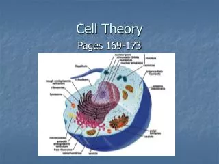
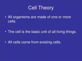
10. The Cell Theory The modern tenets of the Cell Theory include:
All known living things are made up of cells.
The cell is structural & functional unit of all living things.
All cells come from pre-existing cells by division.
(Spontaneous Generation does not occur).
Cells contains hereditary information which is passed from cell to cell during cell division. (The first cell is the exception because it could not have come from a previously existing cell)
All cells are basically the same in chemical composition.
All energy flow (metabolism & biochemistry) of life occurs within cells.
The Cell Theory is to Biology as Atomic Theory is to
Physics.
11. Journal Entry
What are the implications of the cell theory?
Due: __________
12. Microscopes That gizmo pictured to the left is a BIG deal. It literally opened up worlds of organisms and information to scientists. It's importance in the history of medicine and our understanding of disease should not be underestimated.
That gizmo is a compound light microscope.
For you, the biology student, it is perhaps the most important tool for you to understand. . .
In learning about the cell the microscope is the most important tool for allowing us to learn about the cell as a whole and the organelles within it.
14. Light Microscope lab In Pairs of two, examine your compound light microscope.
Label all the parts of your compound light microscope on the sheet provided to you.
You will be quizzed on the structure, function and uses of the compound microscope.
15. E- Microscope Lab Lab on the use of the microscope
Lab pairs of two.
Complete Lab Activity Sheet.