Ear: Your Sensory Organ for Hearing & Equilibrium
Explore the intricate components of the ear - external, middle, and inner - and learn about their functions in hearing and maintaining balance, as well as common hearing loss types, equilibrium disorders, and assessment techniques. Discover how the ear processes sound waves and converts them into electrical impulses for interpretation by the brain.

Ear: Your Sensory Organ for Hearing & Equilibrium
E N D
Presentation Transcript
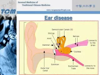
EAR • The sensory organ for hearing & maintaining equilibrium • Has 3 parts: • 1- external (auricle or pinna) • 2-middle • 3- Inner.
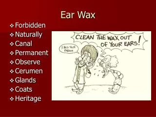
** External: • Serves to funnel sound waves inside, external auditory canal terminates at eardrum (tympanic membrane), canal lined with glands secrete cerumen (yellow waxy material lubricate & protect ear. • Eardrum separate external & middle ear, pulled in by malleus.
** Middle: • Air-filled cavity inside temporal bone • Contains malleous, incus& stapes, open into inner ear, another opening is Eustachian tube → connects middle ear with nasophaynx opens with swallowing or yawning.
3 functions 1- conduct sound vibrations from outer ear to central hearing apparatus in inner ear. 2- protect inner ear by reducing amplitude of loud sounds 3- Eustachian tube allows equalization of air pressure on each side of tympanic membrane so doesn’t rupture.
** Inner: • Bony labyrinth, which holds sensory organs for equilibrium & hearing • Vestibule & semicircular canal (Vestibular apparatus), & cochlea contains central hearing apparatus
Hearing: • Auditory system divided into 3 levels: peripheral, brainstem& cerebral cortex. • Peripheral → ear transmits sounds & converts its vibrations into electrical impulses analyzed by brain. Amplitude (how loud)& frequency (pitch). • Basilar membrane vibrates at specific frequency of sound; mediate vibrations into electric impulses conducted by CN VIII to brainstem.
Hearing: cont Brainstem function is binaural interaction → permits locating direction of a sound in space as well as identifying the sound. Timing of messages from 2 ears depending on the way the head is turned, then cortex interpret the meaning of sound & give response in less than a second.
Pathways of hearing: • Normally its air conducted (AC) as mentioned before alternate is bone conducted (BC) → bones of skull vibrated then transmitted to inner ear & CN VIII
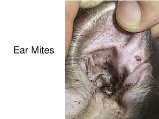
Hearing Loss: • What obstruct sound transition impair hearing • a conductive hearing loss involves a mechanical dysfunction of external & middle ear, but partial (able to hear if amplitude increased enough to reach normal nerve elements in inner ear). • Causes of conductive hearing loss → impacted cerumen, foreign bodies, perforated eardrum, and pus in middle ear.
Sensorineural: • Perceptive loss (pathology of inner ear, CN VIII, auditory areas of cerebral cortex.) • OR caused by presbycusis → gradual nerve degeneration with aging & by ototoxic drugs. • Mixed loss → combinations of conductive & sensorineural types in same ear.
Equilibrium: • Labyrinth feeds information to brain about body's position in space in relation to gravity. • If labyrinth inflamed →wrong information →staggering gait& whirling sensation (vertigo).
** subjective Data: • 1-Earache • 2- infections • 3- discharge • 4- hearing loss • 5- environmental noise • 6- tinnitus • 7- vertigo • 8- self-care behaviors
** Objective data: - Position adult sitting up straight with head at eye level.
Inspect & Palpate External Ear • Size& shape: ears are of equal size bilaterally with no swelling or thickening, taking in consideration a normal familial trait. • Skin condition: skin color is consistent with facial skin color; skin is intact, with no lump or lesions. May note Darwin’s tubercle (small painless nodule at the helix which is congenital variation& not significant) • Tenderness: move the pinna & push the tragus, should feel firm with no pain during movement. Palpate mastoid process, no pain
- External auditory meatus: • no swelling, redness, or discharge should be present. Not size of the opening to direct your choice of speculum for the Otoscope. Some cerumen is usually present (color varies from gray yellow to light brown & black, its texture varies from moist & waxy to dry desiccated)
Inspect Using Otoscope: • Inspect external ear canal noting size of the auditory meatus; choose the largest speculum that will fit comfortably in the ear canal. Tilt person’s head slightly away from you toward the opposite shoulder for better view. • Note any redness & swelling, lesions, foreign body, or discharge. • Pull pinna up & back on adult to straighten S shape of canal , for infant & children less than 3 years pull pinna down ward.
Inspect Using Otoscope: cont • Hold Otoscope “upside down” & have the dorsa of hand along his cheeks. • Insert speculum slowly into canal, put your eye up to Otoscope towards nose may rotate, Otoscope exam before hearing test.
- Tympanic membrane: • Color & characteristics: normal eardrum is shiny & translucent, with a pearl-gray color. • Cone –shaped light reflex: is prominent in the anterior inferior quadrant (at 5 o’clock in rt drum, & 7 o’clock in lt drum). • Sections of malleus, umbo, manubrium, & short process are visible; infrequently incus may be seen behind the drum.
- Tympanic membrane: cont • Position: eardrum is flat, slightly pulled in the center, & flutters when person perform valsalva maneuver (avoid with aging person → may disrupt equilibrium &upper respiratory infections→ may propel infections into middle ear.) • Integrity of membrane: intact eardrum
Test hearing acuity: • This begins during history. an audiometer also used to assess ability to hear sounds of varying frequency. Alternate method is “Crude tests” are nonquantitative; do not measure degree of loss, just document presence of hearing loss
* Voice test: • test one ear at a time, placing one finger on tragus, pushing it in & out of auditory meatus. 30-60 cm from his ear, whisper 2 syllable words. * Tuning fork tests: • Test hearing by (AC){more sensitive} in which sound vibrates through ear canal & middle ear, or (BC) sound vibrates through cranial bones to the inner ear. • To activate fork, hold it by stem & strike the tines softly on back of your hand
Weber test: • valuable when person reports hearing better with one ear than the other, place vibrating tuning fork in midline of his skull. Person hears sound by bone conduction through skull & should sound equal loud both ears.
Rinne test: • compare (AC) & (BC) sound. A normal response is a positive Rinne test, or AC > BC. Repeat with other ear. • Place stem of fork on mastoid & ask him to signalwhen sound goes awaythen quickly invert fork so vibrating end is near ear canal, should still hear.
The Vestibular Apparatus: • Assess ability of the Vestibular apparatus in inner ear to maintain standing balance • may occur), stand close to catch person in case of fall.
Romberg test: • ask person to stand up with feet together & arms at the sides. Ask person to close eyes & to hold the position. Wait about 20 seconds. Normally a person can maintain posture & balance even with blocked visual orientation information, (slight swing (swaying)