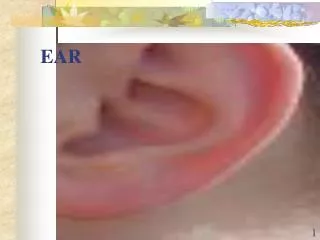
EAR
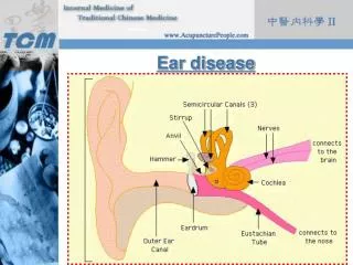
EAR. Outer Ear. Auricle: Layer of skin over cartilage framework. Lobule = fibrous fatty tissue with no cartilage. Sensory innervation: Great auricular nerve. Auriculotemporal nerve. Lesser occipital nerve. Outer Ear. External acoustic (auditory) meatus:

EAR
E N D
Presentation Transcript
Outer Ear • Auricle: Layer of skin over cartilage framework. Lobule = fibrous fatty tissue with no cartilage. • Sensory innervation: Great auricular nerve. Auriculotemporal nerve. Lesser occipital nerve.
Outer Ear • External acoustic (auditory) meatus: From auricle to tympanic membrane. 2.5 cm in length. S-shaped. Lined by skin and ceruminous glands.
Middle Ear • Tympanic membrane: Dense fibrous membrane. Covered by skin externally. Covered by mucosa internally.
Middle Ear • Tympanic cavity: Air-filled cavity within petrous portion of temporal bone. Round window: Enclosed by secondary tympanic membrane: Undergoes compensatory excursions with movement of stapes. Oval window: Closed by footplate of stapes.
Middle Ear • Auditory ossicles: Malleus: Attached to tympanic membrane. Incus. Stapes: Attached to oval window (fenestra vestibuli).
Middle Ear • Stapedius muscle: Inserts onto neck of stapes. Contracts reflexively in response to loud sounds. Innervated by CN VII.
Middle Ear • Tensor tympani muscle: Inserts onto malleus. Contracts reflexively to loud sounds. Innervated by CN V-3.
Middle Ear • CN IX: Tympanic nerve: GSA to mucosa of tympanic cavity, mastoid air cells, and auditory tube. Lateral petrosal nerve: Leaves tympanic cavity to floor of middle cranial fossa. Descends through foramen ovale to infratemporal fossa. Ends in otic ganglion.
Inner Ear • Lies within petrous part of temporal bone. • Consists of: Bony labyrinth: Lined with periosteum. Membranous labyrinth. • Supplied by: Labyrinthine artery (from basilar artery): (Basilar artery is formed by fusion of two vertebral arteries).
Inner Ear • Cochlea: Resembles a snail shell. Three turns. Round window (fenestra cochlea). • Vestibule: Contains: Utricle. Saccule.
Inner Ear • Cochlear (auditory) nerve (CN VIII). • Semicircular canals: Anterior, posterior, lateral. Arranged in three planes. Open into utricle. Dilated at one end to form ampulla: With crista ampullaris. Senses rotational acceleration.
Inner Ear • Utricle and saccule: With macula. Senses linear acceleration and pull of gravity. • Vestibular nerve (CN VIII):