Atomic Force Microscopy (AFM)
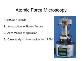
Atomic Force Microscopy (AFM). David Ji Feb 21 06. AFM:. An fancier stylus-based instruments than record players. "Eyes of Nanotechnology". A high-resolution imaging technique that can resolve features as small as an atomic lattice in the real space.


Atomic Force Microscopy (AFM)
E N D
Presentation Transcript
Atomic Force Microscopy (AFM) David Ji Feb 21 06 AFM: An fancier stylus-based instruments than record players "Eyes of Nanotechnology" A high-resolution imaging technique that can resolve features as small as an atomic lattice in the real space. AFM image of A Digital Video Disc Surface
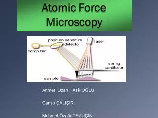
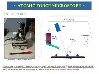
How AFM works? The beam-bounce method 1. Laser 2. Mirror 3. Photodetector 4. Amplifier 5. Register 6. Sample 7. Tip: sharp 8. Cantilever: elastic
How AFM works? Sample under the AFM tip move Force exerted by the tip on the sample is constant. The height changes of the scanner reflect the topography of the sample. Varying force/constant height Piezoelectric crystal: This crystal creates a voltage if pressure is applied, or in reverse, can create a pressure by expanding or contracting if a voltage is applied.
About tips: Higher aspect ratio Tip end radius generally limits the resolution of AFM
Major Modes of Operation Contact Mode Tip scans the sample in close contact with the surface. Cantilever against the sample surface with a piezoelectric positioning element, it is up and down as it scans Non-contact Mode Tip hovers 50-150 Å above a sample surface. Attractive forces acting between the tip and the sample are detected. Tapping Mode (intermittent contact) Tip in contact with the surface and then lifted off the surface to avoid dragging the tip across the surface. the cantilever assembly oscillated at or near its resonant frequency using a piezoelectric crystal. The piezo motion causes the cantilever to oscillate with a high amplitude( typically greater than 20nm) when the tip is not in contact with the surface. The oscillating tip is then moved toward the surface until it begins to lightly touch, or tap.
Summary of imaging modes • Contact mode: Electrostatic and/or surface tension forces from the adsorbed gas layer will pull the scanning tip toward the surface. It can damage samples and distort image data. Contact mode imaging is heavily influenced by frictional and adhesive forces compared to non-contact or tapping mode. • Non-contact mode: Imaging generally provides low resolution and can also be hampered by the contaminant layer which can interfere with oscillation. • Tapping Mode: High resolution without inducing destructive frictional forces both in air and fluid. The very soft and fragile samples can be imaged successfully.
More operation modes Thank you!