Tracheostomy
Tracheostomy. Dr. S. Parthasarathy MD., DA., DNB, MD ( Acu ), Dip. Diab . DCA, Dip. Software statistics PhD ( physio ). Definition . The term tracheotomy refers to the formation of a surgical opening in the trachea . Nomenclature ?? It refers to a temporary procedure.

Tracheostomy
E N D
Presentation Transcript
Tracheostomy Dr. S. Parthasarathy MD., DA., DNB, MD (Acu), Dip. Diab. DCA, Dip. Software statistics PhD (physio)
Definition • The term tracheotomy refers to the formation of a surgical opening in the trachea. • Nomenclature ?? • It refers to a temporary procedure. • Tracheostomyon the other hand refers to the creation of a permanent stoma between the trachea and the cervical skin
History • First described in Rig veda in 2000 BC • In the 1800s tracheostomy gained in popularity, as it became a recognised way of treating patients with Diphtheria. • High and low • Chevaliar Jackson 1909 mortality -- 25% to 1–2% by passage of a few decades
Indications • 1. Mechanical obstruction of the upper airways. • 2. Protection of tracheobronchial tree in patients at risk of aspiration. • 3. Respiratory failure. • 4. Retention of bronchial secretions. • 5. Elective tracheostomy, e.g. during major head and neck surgery a tracheostomy • can provide/improve surgical access and facilitate ventilation.
Mechanical (1) • Congenital : Subglottic or upper tracheal stenosis, laryngeal web, laryngeal and vallecular cysts, tracheo-oesophageal anomalies or haemangioma of the larynx. • Infective Acute epiglottitis, laryngotracheo bronchitis, diphtheria or Ludwig’s angina. • Malignancy Advanced tumours of larynx, tongue, pharynx or upper trachea presenting with stridor. • .
Mechanical (1) • Trauma • Gunshot and knife wounds to the neck, inhalation of steam or smoke, swallowing of corrosive fluid. • Vocal cord paralysis • Post-op complication of thyroidectomy, cardiac or • oesophageal surgery, bulbar palsy. • Foreign body Swallowed or inhaled object lodged in upper airway causing stridor
Neuro • Neurological diseases • GBS, Bulbar palsy,polio,MND • Coma • GCS < 8
RESPIRATORY FAILURE • Pulmonary diseases (exacerbation of chronic bronchitis and emphysema, severe asthma, severe pneumonia). Severe chest injury (flail chest).
RETENTION OF BRONCHIAL SECRETIONS • chronic pulmonary disease, • acute respiratory infection, decreased level of consciousness, and trauma to the thoracic cage or musculature with in-effective cough and retention of secretions
Described originally • But now ---
Recently • 1. Prolonged or expected prolonged intubation • 2. Inability of patient to manage secretions • 3. Facilitation of ventilation support • 4. Inability to intubate • 5. Adjunct to manage head and neck surgery • 6. Adjunct to manage significant head and neck trauma • FESTIVALS - pneumonic
Problems with physiology • warming, humidification and filtering of air do not take place before the inspired air reaches the trachea and lungs. • The normal mucociliary clearance mechanism is disrupted. • Cant talk • Cant swallow with ease • Weak cough reflex
Advantages • Safe and secure • Comfort for the patient • Less sedation needed • Suction easier • Pneumonias less • Resistance is less ( dead space decrease by 70 ml)
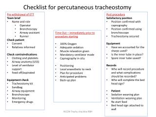
Preparation • Informed consent • Neck examination • Bleeding disorders • A good assistant • Procedure • Anaesthesia or not • Emergency or elective
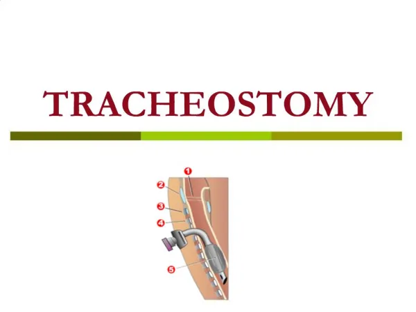
Size !! • The size of the tracheostomy tube usually refers to the inner diameter of the inner cannula. • In other words, the inner diameter reflects the narrowest diameter of a particular tracheostomy tube. • two sizing systems for tracheostomies: the Jackson sizing system and the International Standards Organization (ISO) sizing system
Inner cannula • purpose of the inner cannula is to clear secretions through cleaning or replacement at regular intervals. The standard inner cannula is used most often in the hospital setting because it frequently provides a 15-mm Adapter.
The Obturator • assist with the insertion of the tracheostomy tube. • Typically, it is necessary to remove the inner cannula in order for the obturator to fit within the outer cannula • Keep it in vision
neck flange • The purpose of the neck flange is to stabilize the tracheostomy tube, seating it against the neck and preventing it from migrating inward. • Labels
The Pilot Balloon and the Inflation Line • The Cuff( high pressure, barrel, foam, tear drop) Capillary perfusion pressure is considered to be between 20–30 cm H2O
Types – numerous • 1) Single cannula --- cuffed and uncuffed • 2) Double cannula --- cuffed and uncuffed • 3)Unique a) fenestrated b) talking c) big size
Original - Colour coding of tubes • Orange 6 • Green 7 • White 8 • Blue 9 • Yellow 10
percutaneous dilatational tracheostomy (PDT) is defined as the placement of a tracheostomy tube with the help of one commercially available set using a series of dilators. • The most commonly used PDT kits are available from Cook Critical Care and Sims Portex, which include the single tapered tracheal dilator (Blue Rhino) developed by Ciaglia. • without direct visualization of the trachea.
The pre requisites • PDT necessitates the administration of adequate pain relief, sedation and neuromuscular blockade to the already intubated and mechanically ventilated patient. • Due to this a PDT cannot be used for emergency airway management as in supraglottis or oro facial trauma. • Otherwise indications are similar
Questions about PDT • Patients under the age of 16 years • Obvious deformities of the airway, previous scars from surgeries like a tracheostomysternotomy, neck oedema, gross obesity, a mass (for example a goitre) or a tumour in the neck make it difficult to easily palpate the local landmarks like the cricoid cartilage.
Inability to optimally extend the patient’s neck due to cervical spine trauma or arthritis, the presence of a short neck or due to severe kyphosis. • Haemo dynamically unstable and difficult airways ??
Advantages • Monitors and lines • Time 30 Vs 60 minutes • Cost and theatre personnel • Less trauma– less bleeding and less infection • Decannulation is easier • Scar is less • In MTS, direct cost is less
Nasogastric feed is stopped at least 2 h before the planned time of PDT and the stomach contents emptied just before the actual procedure to prevent aspiration into the airway. • Position similar • 100% oxygen; adequate analgesia, • sedation and muscle relaxation are also ensured
Ventilator settings adjusted for leaks • PEEP 10 – 15 cm • All monitors and IVF and drugs • Bronchoscope introduced • ET tube withdrawn and cuff deflation adequate
The ultimate tube placement is made at the level of the first and the second tracheal cartilages or between the second and the third tracheal cartilages whenever possible. • A 2–3 mm midline subcricoid transverse incision is made on the skin at the pre-selected site.