
Bacteria Staining and Isolation Techniques
E N D
Presentation Transcript
Isolation of Bacteria • Most bacterial samples have numerous different bacterium • Identification requires testing on an individual type of bacteria • Two methods for isolating are the Streak plate and the Spread plate
Streak Plate • Transfer bacteria to a small area of agar plate • Get increasingly smaller amounts of bacteria on successive sections of the plate by sterilizing the loop and spreading the previous area • When you get a small enough quantity of bacteria in an area, they will be able to grow in individual colonies
Streak Plate continued • Objective: isolate single colonies of bacteria from a mixed culture • Draw procedure diagram off of board • Flame loop between each quadrant, but do NOT dip the loop back in the broth tube • Each student will do their own. This is worth 3 points (1 pt labeling, 1 pt technique, 1 pt isolation)
Spread Plate • Transfer a big drop of bacteria to the plate, then spread in all over • Series of dilutions required to get a sample with few enough bacterial cells to produce individual colonies. • We will not perform the dilutions in this lab. We will just learn the spreading technique.
Spread Plate continued • Objective: isolate single colonies of bacteria from a mixed culture (or at least learn the technique that you would use) • After transferring a couple of drops mixed culture to the agar plate, sterilize spreader • Dip in ethanol • Flame (do NOT dip back in ethanol) • Let cool
Stain Categories • Morphological – size, shape, arrangement • Simple stain and Negative stain • Differential – cell wall composition • Gram stain and Acid-fast stain • Structural – cell structures • Endospore stain and Capsule stain
Stains • Basic stains (+) – react with acidic (-) parts of the cell • ex. crystal violet, safranin, methylene blue • i.e. stains that get inside the cell • Acidic stains (-) – are repelled by the negatively charged cell surface • Ex. Congo red and india ink • Stains the background, not the cells
Simple Stain • Objective: Determine morphology and arrangement • All bacteria will be stained • Make a smear prep • Method of getting bacteria adhered to the slide • see next slide for procedure • Each pair of student will do a simple stain on M. luteus
Negative Stain • Objective: Determine morphology and arrangement • India ink used to stain background, not cells • Gives a good view of morphology • Not heat fixed, so cells are not distorted • Prepare negative stain with M. luteus
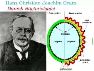
Gram Stain • Objective: determine if bacteria is gram positive or negative • Initial procedure to determine unknown bacteria • Takes advantage of differences in cell wall composition (differential stain) • Gram positive has thick layer of peptidoglycan • Gram negative has thin layer of peptidoglycan
Gram Stain • Gram stain: • E. coli • S. aureus • E.coli/S. aureus mixture • Remember, next week you will be doing a gram stain for points, so get it down today!