BOLD Contrast: Functional Imaging with MRI
BOLD Contrast: Functional Imaging with MRI. Mark A. Elliott, PhD Department of Radiology University of Pennsylvania. Overview. Mechanisms of functional imaging with MRI Methodology of fMRI Issues for animal studies Spatial and temporal sensitivy of fMRI. electrical activity. - excitatory.


BOLD Contrast: Functional Imaging with MRI
E N D
Presentation Transcript
BOLD Contrast:Functional Imaging with MRI Mark A. Elliott, PhD Department of Radiology University of Pennsylvania
Overview • Mechanisms of functional imaging with MRI • Methodology of fMRI • Issues for animal studies • Spatial and temporal sensitivy of fMRI
electrical activity - excitatory - inhibitory - soma action potential electrophysiology Methods for Imaging Neural Activity metabolic response FDG PET - ATP tightly regulated - glucose consumption - oxygen consumption H215O PET hemodynamic response - blood flow fNIR - blood volume - blood oxygenation fMRI EEG MEG Perfusion MRI
Vascular Sensitivity offMRI and fNIR Arterial Venous II I fNIR Intravascular Perfusion MRI II IV fMRI III I Extravascular III IV Vessel Size
Vascular Response fMRI vs fNIR
Mechanisms of fMRI Signal:BOLD Contrast Neural Activity “Flooding the garden to feed the thirsty flower” - ??? CMR02 CBF BOLD ( CBF - CMR02) spatial dimension • Hemodynamic response is a surrogate marker for neural activity • BOLD = Blood Oxygenation Level-Dependent • BOLD signal is a complex interaction of CBF + CBV + CMRO2: • CBF >> CMRO2 less deoxyhemoglobin with activation • CBF is monitored indirectly • “Tracer” is primarily venous • “Tracer” is endogenous
Magnetic Susceptibility Affects Background Magnetic Field : permeability r : relative permeability M: magnetic susceptibility For biological tissues, | M | << 1 Diagmagnetic: M < 0 Paramagnetic: M > 0 The interface between regions with different M behaves like a magnetized dipole, perturbing the local B field. M creates larger B B1 B2 B1 M1 B2 M2
BOLD Contrast: Changes in Magnetic Susceptibility of Blood • Blood and brain tissue are diamagnetic. • Hb0 is diamagnetic. • Hb is strongly paramagnetic. • HbO is paramagnetic. • Increased Neuronal activity: • blood flow increases ≈ 30% • 02 consumption increases ≈ 5% • [Hb0] • [Hb] • Decrease in [Hb] reduces the M between blood and brain tissue • Magnetic field becomes more uniform MRI signal affected
Hypoxia Normoxia Hemoglobin Saturation AffectsMagnetic Field Homogeniety Rat brain, 7T from Ogawa, 1990 Field Map vs. Hemoglobin Saturation from Bandettini and Wong, 1995
Summary: BOLD Contrast in fMRI Verbal Fluency Task Broca’s area Wernicke’s area • BOLD = Blood Oxygenation Level-Dependent • Oversupply of CBF raises [HbO] in regions of increased CMRO2 • Susceptibility mismatch between blood and tissue is reduced • Magnetic field becomes more homogeneous • Temporal T2* contrast generated in T2* sensitive MRI
fMRI Methodology: Acqusition structural T1 weighted ~ 5 min Temporal series of EPIs . . . . 1x1x1 mm voxels EPI functional T2* weighted ~ 2 sec/volume ~ 300 images time ~ 10 min 3x3x3 mm voxels
fMRI Methodology: Stimulus Blocked Design, Event-Related Design, and ISI Fixed ISI ISI On Blocked Design Off On Event Related Off Event related designs can have either fixed or variableinter-stimulus interval (ISI) ISI Variable ISI • Variable ISI allows for more stimuli per time. • Increased statistical power in analysis.
fMRI Methodology: Analysis “Non-Activation” Signal Stimulus . . . . Processing “Activation” Signal Processing Stimulus
T2*-weighted Snapshot Image Average Difference Image Statistical Significance Image Thresholded Statistical Image Overlay on Anatomic Image Brain Activation MapsStatistical Parametric Mapping ON task OFF signal courtesy J. Detre
Hemodynamic Response Function The “HRF” - the theoretical impulse response of BOLD contrast to brief neuronal activity Peak Contrast Amplitude FWHM Onset Time Stimulus Time to Peak
fMRI Model: HRF Linear System Linear Model Assumption y = hx Expected signal (y) is convolution of the stimulus signal (x) with the HRF (h) stimulus (x) HRF (h) signal (y) Signal is predicted for any arbitrary sequence of stimuli
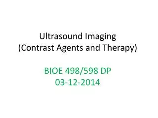
Applications of fMRI • Cognitive Neuroscience • Localization of sensorimotor and cognitive function • Brain-behavior correlations • Clinical Neuroscience • Presurgical mapping • Differential diagnosis of cognitive disorders • Recovery of function/neuroplasticity Photic Stimulation
Implications for Animal fMRI • Pharmacological effects on neuronal metabolism and hemodynamic response • Small voxel sizes reduce SNR • Smaller volumes enable higher field magnets (7 and 9.4T) • Passive stimulus delivery (training possible in some models)
T2* Signal Loss in the Pre-Frontal Cortex Air is highly paramagnetic (like Hb) Air-tissue interface has “static” M Background signal “drop-out” F B0 E 1 S B0 2 Bn = Normal component Bn = Tangential component F = frontal sinus E = ethmoidal sinus S = sphenoidal sinus Normal component is unchanged by B1n = B2n Tangential component is altered by B1n = 1 / 2 B2n
Signal Dropout in T2* Weighted Images Increasing TE
Spatial Extent of BOLD Neural Activity CMR02 Hb Saturation (%, approx.) restingactive arterioles 90 90 capillaries 80 90 veins 60 90 CBF BOLD ( CBF - CMR02) microvessels draining veins Positive T2* contrast derived from CBF > CMR02 Venous compartment experiences largest Hb (and T2*) Draining veins are less spatially specific to site of neural activity
Extravascular BOLD Signal B0 “inhomogeneity” from vessel extends into extravascular (EV) space EV Magnetic Field Gradient microvessel macrovessel • Diffusion of water molecules through B0 gradients • Large vessels: static dephasing, T2* effect • Small vessels: dynamic dephasing, T2 and T2* effect • Spin-echo fMRI less sensitive to large vessel (venous) • extravascular space water diffusion from Principles of Functional MRI, Seong-Gi Kim
Echo Time and Field Strength Effects on BOLD Contrast • BOLD contrast increase with echo time (TE) • SNR decreases with echo time • Optimal CNR when TE resting T2* • BOLD contrast increases with magnetic field • SNR increases with magnetic field 1.5T % Signal TE (msec) 3T % Signal TE (msec) from Stroman et al, Proc. ISMRM, Glasgow (2001)
Field Strength Effecton BOLD Spatial Sensitivity • T2* of blood shortens quadratically with B0 • Field dependence of T2,blood T2,tissue • - Decreased venous contribution Diffusion weighted BOLD Intravascular BOLD component model simulation Rat brain, 9.4T from S.P. Lee et al, (2003)