ORAL MUCOSA
ORAL MUCOSA. 口腔黏膜. 王文岑 助理教授 高醫大附設醫院 S 棟 2 樓 口腔病理影像診斷科 07-3208284 wcwang@kmu.edu.tw . 學習目標. 口腔黏膜的分類 、 定義與功能 口腔黏膜的細胞組成及顯微構造 學習資源及主要圖片引用 : 1. Nanci A: Ten Cate’s Oral Histology, Development, structure, and function 6th ed. p.329-75


ORAL MUCOSA
E N D
Presentation Transcript
ORAL MUCOSA 口腔黏膜 王文岑 助理教授 高醫大附設醫院S 棟 2 樓 口腔病理影像診斷科 07-3208284 wcwang@kmu.edu.tw
學習目標 • 口腔黏膜的分類、定義與功能 • 口腔黏膜的細胞組成及顯微構造 學習資源及主要圖片引用: 1. NanciA: Ten Cate’s Oral Histology, Development, structure, and function 6th ed. p.329-75 2. Orban BJ :Orban’s oral histology and embryology,9th ed. p.261-334 3. Avery JK: Essentials of Oral Histology and Embryology: A clinical approach. 2nd ed. p.164-83
Oral mucosa MUCOUS MEMBRANE Definition: -Moist lining of the intestinal tract, nasal passages and other body cavities that communicate with the exterior Oral mucosa: Oral mucous membrane
各部位的口腔黏膜 上下唇- 唇紅緣 vermillion border 唇黏膜 labial mucosa 唇繫帶 labial frenum 前庭區 vestibule
頰- 頰黏膜 buccal mucosa 頰繫帶 buccal frenum 咬合線 occlusal line 腮腺開口 orifice of Stenson’s duct
舌- 口底- 舌背 唾液腺開口 dorsal surface 舌腹 ventral surfrace 舌繫帶 lingual frenum 舌側緣 tongue border
牙齦- 齒間牙齦 interdental papilla 固著牙齦 attached gingiva 黏膜牙齦 mucogingiva 前庭區
顎部- 硬顎 hard palate 軟顎 soft paalte 懸雍垂 uvula
Oral mucosa FUNCTIONS OF ORAL MUCOSA • Protection • Sensation • Secretion • Thermal regulation
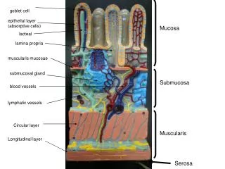
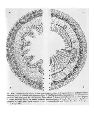
Oral mucosa STRUCTURE OF ORAL MUCOSA • Epithelium……………… ..epidermis • Lamina propria……… ..dermis • Submucosa…………… ..subcutaneous * Epithelial ridges, rete pegs
Oral mucosa EPITHELIUM …stratified squamous cell Keratinized Nonkeratinized basal basal prickle pricklegranular intermediatekeratinized superficial
Keratinization Basal cell prickle granular keratinized layer
Oral mucosa Basal cell layer • Cuboid or columnar shape • Containing bundles of tonofibrils and cell organs • Synthesize DNA, protein • Most cell divisions
Oral mucosa Prickle cell layer(spinous cell layer) • Irregularly polyhedral shape • Conspicuous tonofibril bundles • Membrane-coating granules in upper part • Intercellular bridge- Desmosome • Most active in protein synthesis
Oral mucosa Granular cell layer • Flatter & wider • Keratohyaline granules • Tonofibrils • Membrane-coating granules • Synthesis protein
Oral mucosa Keratin layer • Flattened & dehydrated • Lost all organelles • Cell filled with packed fibrillar material orthokeratin parakeratin
Oral mucosa Intermediate layer of nonkeratinized epi. • Flattened cells • Tonofilaments & glycogen Superficial layer of nonkeratinized epi. • Flattened cells • Tonofilaments & glycogen • Fewer organells • Nuclei persist
Oral mucosa Nonkeratinocytes in the oral epi. • Melanocyte melanin, premelanosome, melanosome • Langerhan’s cell regulatory, antigen trap • Merkel’s cell tactile sensory • Lymphocyte inflammatory response **Clear cells: Melanocyte, Langerhan’s cell, Merkel’s cell
Oral mucosa Intercellular Junctions • Desmosome (Hemidesmosome) • Tight junction • Gap junction Intercelluar bridge
Oral mucosa FUNCTIONAL CLASSIFICATION OF ORAL MUCOSA Keratinized areas • Masticatory mucosa-hard palate & gingiva • Vermilion border Nonkeratinized areas …Lining or reflecting mucosa • lip, cheek, alveolar mucosa, vestibular fornix, mouth floor, soft palate, ventrum of tongue Specialized mucosa • dorsum of the tongue
Oral mucosa Hard Palate 1.different zones of palatine mucosa • gingival region, palatine raphae, fatty zone, glandular zone(mucous gland)
2. tight fixed to the underlying periosteum * No submucous layer: gingiva region and palatin raphe (mucoperiosteum) 3. epithelium uinform in form, well-keratinized
Oral mucosa Gingiva 1. Parakeratinized 75%, orthokeratinized 15%, nonkeratinized 10% 2. often showing stippled surface
3. Free gingiva, free gingiva groove, attached gingiva, interdental gingiva, mucogingival junction 4. “col”…depressed part of interdental papilla, thin nonkeratinized epi.
Oral mucosa Vermilion border of lip • Transitional zone between skin of lip and lip mucosa • No gland
Oral mucosa Nonkeratinized Areas … Lining or reflecting mucosa • lip,cheek, alveolar mucosa, vestibular fornix, mouth floor, soft palate, ventrum of tongue • Thick nonkeratinized epi. and varied amount of loose textured submucosa (containing fat & gland) movably attached to the deep structure
Oral mucosa Fordyce’s spots (granule) • Isolated sebaceous gland • buccal mucosa near mouth angle and opposite the molars
Oral mucosa Specialized Mucosa … Dorsal lingual mucosa • Lingual Papillae Filiform papilla: entire ant part of tongue, no taste bud Fungiform papilla: tongue tip Foliate papilla: lateral border of post. Tongue
Circumvallate papilla: before V-shape terminal sulcus, 8-10 in number • von Ebner’s glands, main source of salivary lipase
Foliate papilla Circumvallate papilla Fungiform papilla
Oral mucosa Taste Buds 1. Ovoid or barrel-shaped intraepi. organ 2. Taste pore 3. Supporting cells 4. 10-12 neuroepithelial cells (receptors of taste)
Oral mucosa Taste Sensation • Sweet-fungiform papilla • Salty-fungiform • Bitter -circumvallate • Sour- foliate
Summary ORAL MUCOSA • STRUCTURE OF ORAL MUCOSA Keratinized vs. Nonkeratinized Keratinocytes in the oral epi Nonkeratinocytesin the oral epi • FUNCTIONAL CLASSIFICATION Keratinized areas Nonkeratinized areas Specialized mucosa
Salivary gland SALIVARY GLAND 唾 液 腺
學習目標 • 唾液腺的分類、組織與細胞組成及功能 學習資源及主要圖片引用: • Avery JK. Oral development and histology. 2nd edition, Chapter 21, p. 352-81 • Berkovitz BK, Holland GR, Moxham BH. 2nd edition, p.215, 220 • de Almeida PDV, et al.. Saliva Composition and Functions: A Comprehensive Review. J Contemp Dent Pract 2008 3:72-80
Salivary gland CLASSIFICATION OF SALIVARY GLAND 1. By location 2. By size Major Salivary Gland • Parotid gland • Submandibular gl. • Sublingual gland Minor Salivary Gland • Labial and buccal, palatine, lingual……
Salivary gland CLASSIFICATION OF SALIVARY GLAND 3. By structure: • Mucous gland • Serous gland • Mixed gland
Salivary gland STRUCTURE OF SG • Acinus cells • Duct system • Connective tissue: • fibrous septa and capsule • blood vessels • nerves
acinic cells mucous cell , serous cell or mixed acini Terminal tubule: acinic cell + myoepithelial cell Terminal secretory units: Terminal tubule+ intercalated duct cell
Salivary gland Mucous cell • pale • Low-protein, high carbohydrate • Mucin: glycoprotein, sialic acid • Viscous • Lubrication
Salivary gland Serous cell • Dark stain • High -protein, low carbohydrate • rER, lysosome, mitochondria, secretory granule, zymogen granules (amylase) • Watery consistency • Digestion
Salivary gland Myoepithelial cell • Surrounding the acinic cell and intercalated duct • Long process • like smooth m. in ultrastructure • Contractile function, helping to extraction
Salivary gland • Demilunes: mixed acini mucous cells (inner) + serous cells • Cell Junctions: • Tight junction • Intermediate junction • Desmosome characteristics of ectodemal origin
Salivary gland SALIVA produced by terminal secretory units striated duct interlobular duct excretory duct main duct (ex. Stensen’s duct) ORAL CAVITY
Salivary gland Parotid gland • Size largest • Location retromandibular fossa, infra-auricular • Main duct Stensen’s duct • Opening buccal mucosa respect to 2nd molar • Acini serous acini • Saliva Vol. 25-30%
Salivary gland Submandibular gland • Size intermediate • Location submandibular triangle, below mylohyoid m. • Main duct Warton’s duct • Opening Caruncula sublingualis, mouth floor • Acini mixed acini, serous predominate • Saliva Vol. 60%