Download
1 / 39
400 likes | 447 Vues
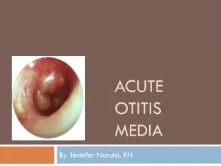
The complications of acute and chronic otitis media. Prof. Abdulrahman Alsanosi Program Director, KSU Fellowship in ORL & Oto-Neurotology Department of Otolaryngology ,Head and Neck surgery King Abdulaziz University Hospital King Saud University. Objectives.

E N D
The complications of acute and chronic otitis media Prof. Abdulrahman Alsanosi Program Director, KSU Fellowship in ORL & Oto-Neurotology Department of Otolaryngology ,Head and Neck surgery King Abdulaziz University Hospital King Saud University
Objectives • The predisposing factors for complications • The pathways for spreading the infections beyond the ear? • To know the classifications of complications • To know presentations ,clinical findings ,investigations and management of each complication
Predisposing factors • Virulent organisms. • Chronicity of disease •Presence of Cholesteatoma and bone erosion. • Obstruction of natural drainage e.g. by a polyp. • Low resistance of the patient
Pathways of infection • extension of infection is by bone erosion due to a cholesteatoma. • Vascular extension (retrograde thrombophlebitis). • Congenital dehiscence • fracture lines • Round or oval window membrane to the labyrinth • Dehiscence due to previous surgery
Classification • Intra-cranial complications • Intratemporal complications • Extra-cranial complications
Intra-cranial complications • Extradural Abscess • Subdural Abscess • Meningitis • Venous Sinus Thrombosis • Brain Abscess
What are the natural barriers between brain and temporal bone ? • Bone • Meninges
Extradural Abscess • Collection of pus against the dura • middle or posterior cranial fossa. • Extradural abscess is the commonest intracranial complication of otitis media
Extradural abscess Clinical Picture – Persistent headache on the side of otitis media. – Pulsating discharge. – Fever – Asymptomatic (discovered during surgery)
Extradural abscess Diagnosis – CT scans reveal the abscess as well as the middle ear pathology. Treatment: – Mastoidectomy and drainage of the abscess
Subdural Abscess Definition – Collection of pus between the dura and the arachnoid. – It’s a rare pathology Clinical picture: – Headache without signs of meningeal irritation – Convulsions – Focal neurological deficit (paralysis, loss of sensation, visual field defects)
Subdural Abscess Investigations – CT scan, MRI Treatment: – Drainage (neurosurgeons) – Systemic antibiotics – Mastoidectomy
Meningitis Definition – Inflammation of meninges (pia & arachinoid) Pathology: – Occurs during acute exacerbation of chronic unsafe middle ear infection.
Meningitis Clinical picture: – General symptoms and signs: • high fever, restlessness, irritability, • photophobia, and delirium. – Signs of meningeal irritation?
Meningitis Diagnosis • – Lumbar puncture is diagnostic: Treatment: – Treatment of the complication itself and control of ear infection: • Specific antibiotics. • Antipyretics and supportive measures • Mastoidectomy to control the ear infection.
Venous Sinus Thrombosis Definition • Thrombophlebitis of the venous sinus. • Etiology: • It usually develops secondary to direct extension
Venous Sinus Thrombosis Clinical picture: – Signs of blood invasion: • (spiking) fever with rigors and chills • persistent fever (septicemia). – Positive Greissinger’s sign which is edema and tenderness over the area of the mastoid emissary Vein. • headache, vomiting, and papilledema(increase intracranial pressure )
Venous Sinus Thrombosis Diagnosis • – CT scan with contrast • – MRI, MRA, MRV • – Angiography, venography • – Blood cultures is positive during the febrile phase.
Venous Sinus Thrombosis Treatment – Medical: • Antibiotics and supportive treatment. • Anticoagulants – Surgical: • Mastoidectomy with exposure of the affected sinus and the intra-sinus abscess is drained.
Brain Abscess Definition • – Localized suppuration in the brain substance. • – It is most lethal complication of suppurative otitis media • Incidence: • – 50% is Otogenic brain abscess
Brain Abscess Pathology • – Site: Temporal lobe or • Less frequently, in the cerebellum. (more dangerous)
Brain Abscess Brain Abscess Diagnosis • – CT scans. • – MRI
Brain Abscess Brain Abscess Treatment Medical: • Systemic antibiotics. • Measure to decrease intracranial pressure. – Surgical: • Neurosurgical drainage of the abscess . • mastoidectomy operation after subsidence of the acute stage.
Intratemporal complications • Labybrinthitis • Ossicular fixation or erosions • Labyrithine fistula • Facial nerve paralysis • Mastoiditis /mastoid abscess
Labyrinthine fistula • Labyrinthine fistula Definition: • communication between middle and inner ear Atiology : • It is caused by erosion of boney labyrinth due cholesteatoma
Labyrinthine fistula Clinical picture : • Hearing loss • Attack of vertigo mostly during straining ,sneezing and lifting heavy object • Positive fistula test
Labyrinthine fistula Diagnosis: • High index of suspicion • longstanding disease • fistula test • Ct scan of temporal bone Treatment : Mastoidectomy
Facial nerve paralysis • Congenital or acquired dehiscence of nerve canal • It is possibly a result of the inflammatory response within the fallopian canal to the acute or chronic otitis media • Tympanic segment is the most commom site to be involved
Facial nerve paralysis Diagnosis • Clinical • May occur in acute or chronic ottis media • Ct scan
Facial nerve paralysis Treatment : -Acute otitis media and acute mastoiditis (cortical mastoidectomy +ventilation tube) - chronic otitis media with cholestetoma ( mastoidecomy ± facial nerve decompresion )
MASTOIDITIS Definition: • It is the inflammation of mucosal lining of antrumand mastoid air cells system.
Acute Mastoiditis Pathology • Production of pus under tension • Hyperaemic decalcification • Osteoclastic resorption of bony walls
Symptoms: • Earache • Fever • Ear discharge Signs: • Mastoid tenderness • Sagging of posterosuperiormeatal wall • TM perforation • Swelling over mastoid • Hearing loss
Mastoid abscess Investigation : • CT scan temporal bones • Ear swab for culture and sensitiveity
TREATMENT Medical treatment: − Hospitalize − Antibiotics − Analgesics Surgical treatment: −Myringotomy − Cortical mastoidectomy
Extracranial complications • Extension of infection to the neck • Bezold abscess ( extension of infection from mastoid to SCM)