A Comparative Study of Prokaryotic & Eukaryotic Cells
630 likes | 665 Vues
Explore the differences in cellular structures of prokaryotic and eukaryotic cells, including membrane compositions, functions, external features, and appendages. Understand the roles of various components in cellular mechanisms.

A Comparative Study of Prokaryotic & Eukaryotic Cells
E N D
Presentation Transcript
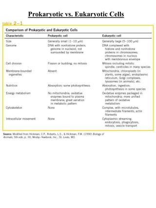
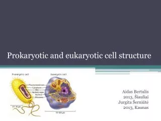
Prokaryotic & Eukaryotic Cells: An Overview • Prokaryotes • Do not have membrane surrounding their DNA • lack a nucleus • Lack various internal structures bound with phospholipid membranes • Are small, ~1.0 µm in diameter • Have a simple structure • Composed of bacteria and archaea
Prokaryotic & Eukaryotic Cells: An Overview • Eukaryotes • Have membrane surrounding their DNA • Have a nucleus • Have internal membrane-bound organelles • Are larger, 10-100 µm in diameter • Have more complex structure • Composed of algae, protozoa, fungi, animals, and plants
Prokaryotic & Eukaryotic Cells: An Overview [INSERT FIGURE 3.1]
Prokaryotic Cell Membrane • Structure • Referred to as phospholipid bilayer; composed of lipids and associated proteins • Approximately half composed of proteins that act as recognition proteins, enzymes, receptors, carriers, or channels • Integral proteins • Peripheral proteins • Glycoproteins • Fluid mosaic model describes current understanding of membrane structure
Cell Membrane Membranes contain a hydrophilic and hydrophobic side Composed of many different types of proteins Proteins in the lipid bilayer move freely within the membrane
Cell Membrane Thin pliable lipid and protein envelope that defines a cell. Phospholipid bilayer Functions: • Regulates nutrient and water intake • Regulates waste removal • Site of prokaryotic respiration • Site of prokaryotic flagella attachment • Involved in the distribution of genetic material during binary fission
Prokaryotic Cytoplasmic Membranes • Function • Energy storage • Harvest light energy in photosynthetic prokaryotes • Selectively permeable • Naturally impermeable to most substances • Proteins allow substances to cross membrane • Occurs by passive or active processes • Maintain concentration and electrical gradient • Chemicals concentrated on one side of the membrane or the other • Voltage exists across the membrane
External Structures of Prokaryotic Cells • Glycocalyces • Gelatinous, sticky substance surrounding the outside of the cell • Composed of polysaccharides, polypeptides, or both
External Structures of Prokaryotic Cells • Types of Glycocalyces • Capsule • Composed of organized repeating units of organic chemicals • Firmly attached to cell surface • Protects cells from drying out • May prevent bacteria from being recognized and destroyed by host
Capsule Polysaccharides or polypeptides in composition. Surround the cell wall in some bacteria. Function: Protection from phagocytosis Osmotic barrier Reservoir for nutrients Virulence factor
Slime Layer Consist of polysaccharide fibers that extend form the bacterial surface Functions: Protection Attachment Associated with biofilms
External Structures of Prokaryotic Cells • Types of Glycocalyces • Slime layer • Loosely attached to cell surface • Water soluble • Protects cells from drying out • Sticky layer that allows prokaryotes to attach to surfaces
Bacterial Appendages Flagella Axial Filaments Pili (Fimbriae)
Bacterial Appendages Structures of locomotion Originate in the plasma membrane In bacteria rotate like a propellar Many different arrangements Flagella
External Structures of Prokaryotic Cells • Flagella • Are responsible for movement • Have long structures that extend beyond cell surface • Are not present on all prokaryotes
External Structures of Prokaryotic Cells • Flagella • Structure • Composed of filament, hook, and basal body • Flagellin protein (filament) deposited in a helix at the lengthening tip • Base of filament inserts into hook • Basal body anchors filament and hook to cell wall by a rod and a series of either two or four rings of integral proteins • Filament capable of rotating 360º
Bacterial Appendages Monotrichous Lophotrichous Amphitrichous Peritrichous Arrangements of Flagella
Bacterial Appendages Axial filament (endoflagella) Originates in the cell membrane and transverses the length of the cell in the periplasmic space. As the endoflagella rotate to move the cell the characteristic shape is formed . Endoflagella are associated with spirochetes.
External Structures of Prokaryotic Cells Endoflagellum is also know as an axial filament. Attached to the plasma embrane and transverses the entire cell. Responsible for the spirochete morphology.
External Structures of Prokaryotic Cells • Flagella • Function • Rotation propels bacterium through environment • Rotation reversible, can be clockwise or counterclockwise • Bacteria move in response to stimuli (taxis) • Runs • Tumbles
Bacterial Appendages • Fimbriae and Pili • Rod-like proteinaceous extensions
Bacterial Appendages Hollow tubes that protrude from some bacteria Compose of protein Fimbriae
External Structures of Prokaryotic Cells • Fimbriae • Sticky, bristlelike projections • Used by bacteria to adhere to one another, to hosts, and to substances in environment • Shorter than flagella • May be hundreds per cell • Serve an important function in biofilms • Virulence factor
External Structures of Prokaryotic Cells • Pili • Tubules composed of pilin • Also known as conjugation pili • Longer than fimbriae but shorter than flagella • Bacteria typically only have one or two per cell • Mediate the transfer of DNA from one cell to another (conjugation)
Transfer of plasmid DNA from a donor to a recipient. Process strengthens the bacterial cell and alows for survival in a competitive environment. Bacterial Conjugation
Bacterial Inclusion Bodies 1. poly-Beta-hydroxybutyric acid - stores lipids for use in plasma membrane 2. glycogen - stores starch like polymer of sugar for energy production 3. Polyphosphate granules (metachromatic granules) - storage for phosphates for plasma membrane and the formation of ATP from ADP. 4. Sulfur granules - stores sulfur which is necessary for the metabolic reactions in biosynthesis.
5. Mesosome Mesosomes - invagination of the plasma membrane that increases the surfaces area of the plasma membrane during binary fission. The mesosome also serves as a site for the attachment and distribution of genetic material during binary fission.
Mesosome In prokaryotic cell division, called binary fission. A diagram of the attachment of bacterial chromosomes, indicating the possible role of the mesosome (an inward fold of the cell membrane) in ensuring the distribution of the "chromosomes" in a dividing cell. Upon attachment to the plasma membrane, the DNA replicates and reattaches at separate points. Continued growth of the cell gradually separates the chromosomes and allocates chromosome copies to the two daughter cells.
Inclusion Bodies 6. gas vacuoles - storage of metabolic gases such as methane or hydrogen gas. The gas vacuoles help in the buoyancy of the cell and aids in it motility. 7. ribosomes - responsible for the synthesis of proteins. 8. nucleoid material - the genetic material of bacteria, which usually is balled up in the cell. During binary fission the nucleoid material unravels within the cell in order to be copied and distributed to the daughter cells. 9. Plasmid - small fragments of self-replicating extrachromosomal DNA that codes for the resistance to antibiotics or for the productions of a specific metabolite, i.e. toxins, pigments. These plasmids may be transferred from one bacterial cell to another by the F-pili.
Inclusion Bodies 9. Plasmid - small fragments of self-replicating extrachromosomal DNA that codes for the resistance to antibiotics or for the productions of a specific metabolite, i.e. toxins, pigments. These plasmids may be transferred from one bacterial cell to another by the F-pili.
Inclusion Bodies These plasmids may be transferred from one bacterial cell to another by the F-pili.
Inclusion Bodies • Endospores - a survival mechanism of certain genera of bacteria such as Clostridium and Bacillus. The endospores are composed of a complex of dipicolinc acid and calcium and the function of the endospore is to protect the bacterial chromosome. The endospores are very resistant to heat, desiccation, freezing, and other physical properties such as pesticides, antibiotics, dyes, and acids.
Inclusion Bodies The endospores may remain dormant for many years until the environment becomes suitable to sustain the life of the bacteria. The endospore will then germinate to form an exact copy of the parent cell that produced it.
Eukaryotic Cell Walls & Cytoplasmic Membranes • Fungi, algae, plants, and some protozoa have cell walls but no glycocalyx • Composed of various polysaccharides • Cellulose found in plant cell walls • Fungal cell walls composed of cellulose, chitin, and/or glucomannan • Algal cell walls composed of cellulose, proteins, agar, carrageenan, silicates, algin, calcium carbonate, or a combination of these
Cell Walls Three different types of cell walls and their compositions: Fungal cell walls are composed of cellulose and/or chitin. Plant cell walls are composed of cellulose. Algal cell walls are composed of cellulose, silicon, and calcium carbonate.
Plasma Membrane Consist of a lipid bilayer and associated proteins. The Plasma Membrane of Eukaryotic cells resembles and functions in the same manner as the prokaryotic plasma membrane with the following exceptions; Contains high levels of sterols such as cholesterol. No respiratory enzymes are located in the eukaryotic plasma membrane. Respiration occurs in the mitochondria.
External Structure of Eukaryotic Cells • Glycocalyces • Never as organized as prokaryotic capsules • Help anchor animal cells to each other • Strengthen cell surface • Provide protection against dehydration • Function in cell-to-cell recognition and communication
Eukaryotic Appendages Flagella There are several different arrangements of flagella in eucaryotes. This diagram represents a biflagellated eukaryotic cell. One of the flagella aids in movement laterally and the other aids in up and down movement. The eukaryotic flagella move like a whip. See Flagellar handout.
Eukaryotic Appendages • Flagella • Function • Do not rotate, but undulate rhythmically
Eukaryotic Appendages Cilia Similar to flagella both structurally and functionally but are much shorter and more numerous. Cilia are found peritrichously to the cell. Move in an undulating manner and motility by those organisms with cilia is much more rapid than those with flagella.
Intracellular Structures of Eukaryotic Organisms (organelles) • Membranous Organelles • Nucleus • Often largest organelle in cell • Contains most of the cell’s DNA • Semi-liquid portion called nucleoplasm • One or more nucleoli present in nucleoplasm; RNA synthesized in nucleoli • Nucleoplasm contains chromatin – masses of DNA associated with histones • Surrounded by nuclear envelope – double membrane composed of two phospholipid bilayers • Nuclear envelope contains nuclear pores