Download

1 / 41
410 likes | 426 Vues
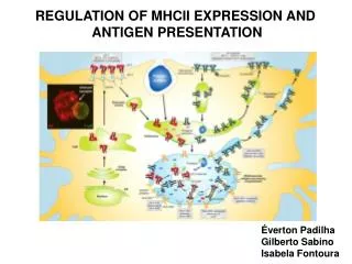
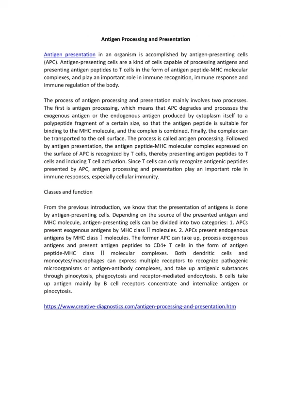
REGULATION OF MHCII expression and antigen presentation. Éverton Padilha Gilberto Sabino Isabela Fontoura. MHCII structure. Expression in: B cells Dendritic cells Macrophages Thymus epitelial cells. Antigen processing and presentation by MHCII. Regulation of MHCII expression.

E N D
REGULATION OF MHCII expressionand antigenpresentation Éverton Padilha Gilberto Sabino Isabela Fontoura
MHCII structure • Expression in: • B cells • Dendritic cells • Macrophages • Thymus epitelial cells
Regulation of MHCII expression CIITA – a master regulation of MHCII expression ( Transactivator of MHCII genes) C-term CIITA gene N-term LRR domain Transcription factors binding Chromatin remodeling Nucleus drive and recruitment of enhanceossome
Gene structure mice Walter Reith et al., 2005. Nature immunology reviews
Pol I Chromatin remodeling Transcription promoter Walter Reith et al., 2005. Nature immunology reviews
Factors that regulates negatively MHCII expression: ↑MARCH-1
Éverton Padilha Role of ubiquitination in pMHCII complex recycling in DCs
What is the role of March-I in pMHC-II ubiquitination and surface expression in both immature and mature DCs?
Validation of methodology C57BL/6 DC with GM-CSF WT KO March-I Knock in MHC II I-Ab immunoprecipitation immunoblotting
How is the expression of MHC class II in immature and mature DC? DC differentiated with GM-CSF WT KO March-I Immature DC Mature DC stimulated with 1mg LPS Facs analysis
DC differentiated with GM-CSF WT KO March-I Immature DC Mature DC stimulated with 1mg LPS Immunofluorescence microscopy How is the intracellular localization of MHC II in mature and immature DCs? Ubiquitination of pMHC-II affects surface expression and intracellular distribution in DCs.
DC with GM-CSF HeLa-CIITA WT KO March-I Knock in MHC II I-Ab Transfected GFP alone GFP+ March I Absence LPS Presence LPS Immunoprecipitation overnight Isotype control IgG Anti-MHC II a-chain mAb The cells were incubated on ice with mAb Y3P Immunoblotting (anti-ubiquitin or anti class II b) Endocytosis were measured for MFI Does ubiquitination be regulating endocytosis in HeLa cells? Does ubiquitination be regulating endocytosis in DCs? The ubiquitination Increase fourfold!
HeLa-CIITA Transfected GFP alone GFP-March I Incubated with mAb against pMHC II Facs analyses Does ubiquitination be regulating endocytosis in HeLa cells? Ubiquitination does not affect the kinetics of pMHC-II endocytosis.
DC with GM-CSF WT DC differentiated with GM-CSF WT KO March-I Knock in MHC II I-Ab Immature DC Mature DC stimulated with 1mg LPS Absence LPS (Immature DC) surface biotinylating The cells were biotinylated on ice. Recultured 8h Immunoprecipitation Immunoprecipitation SDS/Page and avidin SDS/Page and avidin How does March-I regulate the MHCII expression on DC surface? Ubiquitination by March-I regulates pMHC-II degradation.
DC with GM-CSF KO March-I WT Treatment with HEL protein overnight Absence LPS Presence LPS overnight Expression of I-Ak HEL46-61 Analyzed by FACS Does March-I regulate MHCII expression in immature DCs by promoting especific degradation? Ubiquitination regulates degradation of internalized pMHC-II complexes in immature DCs.
Immature dendritic cells Adapted of Lysosome Berger A C , Roche P A J Cell Sci 2009;122:1-4
Goal: Investigate the regulation of MHC class II antigen presentation in three processes: peptide loading, transcriptional regulation, and general cell biology which consists in the assembly, intracellular transport, processing in the MIIC, and endo- and exocytosis.
Fundamental questions… What regulates MHC-II transcription? What controls MHC-II transport in dendritic cells?
Method validation 13 candidates with function already known in literature.
Method validation genetic association 8% (21 of 276) were associated with autoimmune diseases
Which genes do affect MHC-II transcription? MelJuSo siRNA 276 candidates genes qPCR HLA – DR mRNA CIITA mRNA Ii mRNA The silencing of nine candidates proteins may affect transcription of one or several of the tested genes.
Is there any interconnection between these nine candidates? MelJuSo Silencing of each 9 candidates genes qPCR Most of siRNA affected the expression of one or more other candidate genes, suggesting that they act in complex networks.
What is the role for RMND5B? MelJuSo siRMND5B qPCR/confocal TGF-β +/- RMND5B might act as an inhibitor of SMADs by a probably interaction, once RMND5B also translocates to nucleus with SMAD4 upon TGF-βexposure.
What about intracellular distribution of MHC-II? 267 Quantitative microscopy analysis
Quantitative microscopy analysis Nine unrelated candidates proteins might control MHC-II distribution.
Do these genes mimic a mDCs phenotype? siRNA MelJuSo imDCs Confocal microscopy 6 dayson GM-CSF Six candidates proteins might control MHC-II distribution.
MHC-II transport to the plasma membrane Monocytes Confocal microscopy/qPCR imDCs 6 dayson GM-CSF PSD4 is a GEF that may upregulate MHC-II expression by activating ARL14/ARF7.
MHC-II transport to the plasma membrane Monocytes Confocal microscopy/IP and WB imDCs 6 dayson GM-CSF lipid biding-assay Collectively, this reveals part of a pathway where PIP5K1A and PIP3KR2 creates PIPs that are requires for recruitment or activation of GEF PSD4, which activates ARL14/ARF7
MHC-II transport to the plasma membrane ARL14/ARF7 connect to the actin network via ARF7EP-MYO1E to control export of MHC-II
Conclusions Transcriptional networks
Conclusions MHC-II distribution pathway to the plasma membrane