Antigen presentation and antigen presenting cell
460 likes | 1.25k Vues
Antigen presentation and antigen presenting cell. The basic process of immune response. Effector cells. Antigen recognition. Lymphocyte activation. Memory cells. Antigen processing. B. Antigen presentation. Y. antigen processing and presentation.

Antigen presentation and antigen presenting cell
E N D
Presentation Transcript
The basic process of immune response Effector cells Antigen recognition Lymphocyte activation Memory cells
Antigen processing B Antigen presentation Y antigen processing and presentation T cells do not recognize native antigens Recognize antigen that has been degraded into antigenic peptides and displayed with MHC molecules on the cell surface T Y BCR TCR antigenic peptide- MHC native antigen APC (antigen-presenting cell)
CD4 T cell recognize peptide associated with MHC class II molecules • CD8 T cell recognize peptide associated with MHC class I molecules
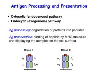
Class I MHC molecule Class II MHC molecule What determines whether an antigenic peptide associates with class I or with class II molecules?
It is dictated by the mode of entry into the cell, either exogenous or endogenous. • Class I MHC molecules bind peptides derived from endogenous antigens • Class II MHC molecules bind peptides derived from exogenous antigens
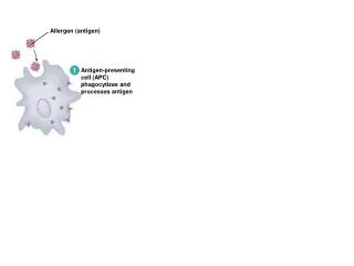
Exogenous antigen-isproduced outside of the host cell enter the cell by endocytosis or phagocytosis
Endogenous antigen-is produced within the host cell itself • viral proteins • tumor proteins
Two processing and presentation pathways • Endogenous antigens • processed in the cytosolicpathway • presented withclass I MHC molecules • Exogenous antigens • processed in the endocytic pathway • presentedwith class II MHC molecules
Endogenous antigens: the cytosolic pathway Intracellular proteins are degraded into short peptidesby a proteasome (a cylindrical particle with a central channel) Degradation of protein occur withinthe central hollow of the proteasome
Newly synthesised MHC class I molecules Peptides need access to the ER in order to be loaded onto MHC class I molecules Peptide antigens produced in the cytoplasm are physically separated from newly formed MHC class I Endoplasmic reticulum (ER) Cytosol
Hydrophobic transmembrane domain Lumen of ER Lumen of ER Peptide Peptide Peptide Peptide Peptide Peptide Peptide Peptide Peptide Peptide Peptide ER membrane ER membrane TAP-1 TAP-1 TAP-1 TAP-1 TAP-1 TAP-1 TAP-1 TAP-1 TAP-1 TAP-1 TAP-1 TAP-2 TAP-2 TAP-2 TAP-2 TAP-2 TAP-2 TAP-2 TAP-2 TAP-2 TAP-2 TAP-2 Cytosol Cytosol ATP-binding cassette (ABC) domain Peptide antigens from proteasome Transporters associated with antigen processing (TAP1 & 2) peptides are translocated by TAP into the ER TAP is optimized to transport peptides thatwill interact with class I MHC
Peptides assemble with class I MHC aided by chaperone molecules Peptide Peptide Peptide Peptide Peptide Peptide Peptide Peptide Peptide Peptide Peptide TAP-1 TAP-1 TAP-1 TAP-1 TAP-1 TAP-1 TAP-2 TAP-2 TAP-2 TAP-2 TAP-2 TAP-2 TAP-2 TAP-2 TAP-2 TAP-2 TAP-2 TAP-1 TAP-1 TAP-1 TAP-1 TAP-1 Endoplasmic reticulum B2-M binds and stabilises floppy MHC Tapasin, calreticulin, TAP form a complex with the floppy MHC Calnexin binds to nascent class I chain until 2-M binds Peptides are loaded onto the MHC molecule and the structure becomes compact
Exported to the cell surface Sent to lysosomes for degradation Fate of MHC class I
Endogenous antigens: the cytosolic pathway Cell surface Golgi ER calnexin Cytoplasm peptide Endogenous antigen
Cell surface Uptake Endosomes Increase in acidity Exogenous antigens: the endocytic pathway degraded into peptides proteases are activated by the decrease in pH Proteases produce 13~18 amino acid long peptides from antigens
MHC class II maturation and invariant chain In the endoplasmic reticulum prevent any endogenously derived peptides from binding to class II molecules invariant chain (Ii chain) interacts with the peptide-binding cleft of the class II molecules
Cell surface Endosomes Uptake MHC class II compartment (M II C) Ii complex exit from the ER a short fragment of the Ii chain termedclass II associated invariant chain peptide (CLIP) occupies the peptide-binding groove
Removal of CLIP ? How can the peptide bind to the peptide-binding groove of class II molecules?
HLA-DM isrequired to catalyze theexchange of CLIP with antigenicpeptides HLA-DM M II C
Transported to the cell surface Sent to lysosomes for degradation Surface expression of MHC class II-peptide complexes
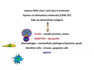
Which cells can present antigen to CD8+ T cells? And Why? • All nucleated cells • Which cells can present antigen to CD4+ T Cells? And Why? • Professional antigen presenting cells Dendritic Cells Macrophages B cells
Antigen presenting cell (APC) • Professional APCs are distinguished by two properties: • express class II MHC molecules on their membranes • deliver a co-stimulatory signal that is necessary for CD4 T cell activation • Nonprofessional APCs: • be induced to express class II MHC molecules or a co-stimulatory signal • function in antigen presentation only for short periods of time during a sustained inflammatory response
Dendritic cells are the most effective of APCs • constitutively express a high level of class II MHC molecules and costimulatory activity • activate naive CD4 T cells • Macrophagesmust be activated before they express class II MHC molecules or the co-stimulatory molecules • B cells constitutively express class II MHC molecules but must be activated before they express the co-stimulatory molecules
Dendritic cell (DC) • acquire their name because of the numerous long membrane extension similar to the dendrites of neurons
Immature vs.Mature DCs Immature DCsexpress TLR, FcgR, C3bR, and class I and II MHC I/II molecules. They are good at antigen uptaking and processing, but poor at inducing T cell activation. Mature DCsare derived from immature DCs upon stimulation by TLR signals and various cytokines, expressing high levels of co-stimulatory molecules. While losing antigen capturing capacity, they are stronger inducers of T activation.
DCs in non-lymphoid tissues Interstitial DC Identified in all tissues other than the brain Langerhans Cell Found in the epidermal layers of the skin Both are particularly good at antigen capturing Nameby Tissue Distribution
DCs in lymphoid tissues Interdigitating DC (IDC) Mainly found in T cell area in lymphoid tissues Expressing high levels of class II MHC and co-stimulatory molecules Particularly potent in T cell activation Follicular DC (FDC) Mainly found in lymphoid follicules Expressing high levels of Fc receptor and complement receptor Critical in germinal center reaction do not functionas antigen presenting cells Name by Tissue Distribution
What to remember The two major pathways for antigen presentation The features of three major types of professional APCs
Study question • Define the following terms: • Exogenous antigen • -isproduced outside of the host cell • Endogenous antigen • -is produced within the host cell itself
Study question • CD4 Th cells recognize antigen with class (II) MHC molecules on (antigen-processing) cells. • CD8 TC cells recognize antigen with class (I) MHC molecules on (target) cells. • (Endogenous) antigens are degraded into peptides within the cytosol by (proteasomes) and assemble with class (I) molecules in the RER.
Study question • Cells that can present antigen to TH cells have been classified into two groups—(professional) and (nonprofessional) APCs. • (Dendritic cells) are the most effective of APCs
Study question • Fill in the blanks in the following statements with the most appropriate terms: a. ( ), ( ) and ( ) all function as professional antigen presenting cells. b. Only antigen-presenting cells express class ( ) MHC molecules, whereas nearly all cells express class ( ) MHC molecules. c. ( ) antigens are internalized by antigen-presenting cells, degraded in the ( ), and displayed with class ( ) MHC molecules on the cell surface. d. ( ) antigens are produced in altered self-cells, degraded in the ( ), and displayed with class ( ) MHC molecules on the cell surface.