Download
1 / 15
170 likes | 352 Vues
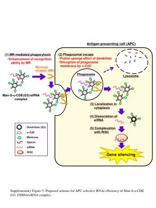
PROFESSIONAL ANTIGEN PRESENTING CELLS. Express MHC class I and class II molecules Express co-stimulatory molecules (CD40, B7) Take up extracellular antigens B cells – soluble proteins , toxins ADAPTIVE – Ag specific Macrophages – extracellular pathogens ( bacteria , yeast )

E N D
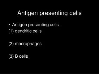
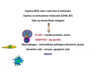
PROFESSIONAL ANTIGEN PRESENTING CELLS Express MHC class I and class II molecules Express co-stimulatorymolecules (CD40, B7) Takeupextracellularantigens B cells – solubleproteins, toxins ADAPTIVE – Agspecific Macrophages – extracellularpathogens (bacteria, yeast) Dendriticcells – viruses, apoptoticcells INNATE
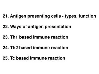
THE ROLE OF PROFESSIONAL ANTIGEN PRESENTING CELLS IN THE IMMUNE RESPONSE Infectiousdiseases Tissuetransplantation Elimination of tumors Autoimmunediseases Gatekeeperfunction Sensingpathogens Primingadaptiveimmuneresponses Maintenance of selftolerancetoselfstructures
Dendriticcellsaresensors, gatekeepers and messengers Activationinducesa phenotypeessentialfor theinitiationof theadaptiveimmuneresponse
CONTACT OF DENDRITIC CELLS AND T - LYMPHOCYTES IN LYMPHOID ORGANS Activateddendriticcellsactasprofessionalantigenpresentingcells MHC-peptidecomplexes1. signal STRANGER Co-stimulatorymolecule2. signal AMPLIFICATION Cytokines3. signal DANGER Theyareinclosecontactwith specific T lymphocytes
INTERDIGITATING RETICULAR (MATURE DENDRITIC) CELL IN T CELL AREAS OF LYMPH NODES NUCLEUS T CELL T CELL CYTOPLASM
Cell-surface molecules of the immunoglobulin superfamily initiate lymphocyte adhesion to professional antigen-presenting cells B. Transient interactions are stabilized by Ag-binding A. Initial contact A
CHANGES OF TISSUE ENVIRONMENT INDUCES THE ACTIVATION OF MACROPHAGES AND DENDRITIC CELLS Phagocytosis and degradation of backteria (LPS, TLR) DANGER SIGNAL Macrophage Activated macrophage Monocyte Dendritic cell Activated dendritic cell Virus, extracellular pathogens, inflammatory cytokines (LPS, TLR) DANGER SIGNAL BLOOD TISSUE LYMPHOID TISSUE
Effector and memory T cells Lymphatics Activated DC Inflammation Pathogen Naive T cells ANTIGEN CIRCULATION Tissue DC ACTIVATION AND MIGRATION OF DENDRITIC CELLS TISSUE LYMPH NODE TISSUE DC AND T CELLS ENCOUNTER T CELL ACTIVATION
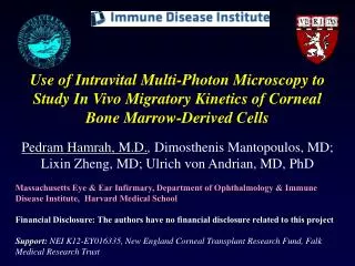
Capture of an Ag-Specific T Cell by an Ag-Bearing DC Rapid DC Migration in the Subcapsular Space Bone-marrow derived DCs (either 5 µM CFSE, green) or (50 µM Cell Tracker Blue, blue) were injected into the footpad of a C57BL/6 mouse, followed 18 hours later by intravenous injection of freshly isolated polyclonal CD4+ T cells (5 µM SNARF, red) and CD8+ T cells (5 µM CFSE and 5 µM SNARF, yellow). The draining LN was removed 6 hours after injection Bone-marrow derived DCs (yellow) were pulsed with 1 µM Ova 4 peptide and 10 µM Ova for 1 hour at 37oC, then injected into the footpad of a C57BL/6 recipient. This was followed 6 hours later by i.v. co-injection of OT-I CD8+ T cells (5 µM CFSE, green) and OT-II CD4+ T cells (5 µM SNARF, red). Huang et al Immunity 2004
Morphology of plasmacytoid dendritic cells IPC/DC2 pDC monocyte Scanning EM Transmission EM
Plasmacytoid DCs control the function of many immunocytes IFNα is impotant in SLE pathology HIV infects PDC Role in immune response and in the pathogenesis of autoimmune diseases and cancer
PLASMACYTOID DENDRITIC CELLS AS PROFESSIONAL TYPE I INTERFERON SECRETING CELLS Enhanced NK cell cytotoxic activity TLR4 Vírus infection TRAM TRIF TLR7 TLR8 TLR9 TLR3 TRIF MyD88 TANK IRAK-1 Activation of and γδ T cells TRAF-6 RIG-1 IKKε TBK1 IFN-β IFN-α1 Cross-presentation by conventional dendritic cells is enhanced IRF-3 IRF-5 IRF-7 IRF-7 Ig production by B cells is induced Type I interferon receptor
Migration Pathways of PDC/IPC versus mDC into a lymph node mDC: afferent lymphatics IPC: HEV Both migrate into the T-cell rich areas