Download
1 / 48
540 likes | 1.07k Vues
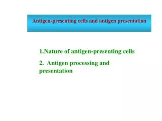
Antigen presenting cells and antigen presentation. B. B. B. B. B. B. B. B. B. Y. Y. Y. Y. Y. Y. Y. Y. Y. Y. T. T. Y. Y. T cells do not recognise native antigens. Y. Y. Y. Y. Y. Y. BCR 交联. 活化增殖、产生抗体. 无增殖 无 CK 产生. Processing and presentation of antigens.

E N D
B B B B B B B B B Y Y Y Y Y Y Y Y Y Y T T Y Y T cells do not recognise native antigens Y Y Y Y Y Y BCR交联 活化增殖、产生抗体 无增殖 无CK产生
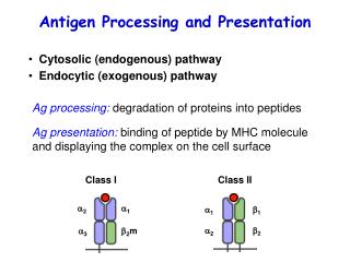
Processing and presentation of antigens • I. APC (antigen presenting cells) • II. Processing and presenting pathway
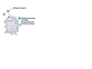
I. Antigen presenting cells • Concepts Endogenous antigens: antigens produced within cells Exogenous antigens: antigens internalized by endocytosis Ag capturing-------Endocytosis Phagocytosis Pinocytosis Receptor-mediated endocytosis Ag processing and Ag presentation • APC
I. Antigen presenting cells • Concepts Endogenous antigens: antigens produced within cells Exogenous antigens: antigens internalized by endocytosis Ag capturing-------Endocytosis Phagocytosis Pinocytosis Receptor-mediated endocytosis Ag processing and Ag presentation A protein antigen be degraded into peptides by a sequence of events The degraded peptides associate with MHC molecules, and the peptides-MHC molecule complexes are transported to the membrane, where they are displayed.
The process of immune response • Exogenous antigens Site of infection peripheral lymphoid organ Peripheral tissue • Endogenous antigens Site of infection peripheral lymphoid organ Peripheral tissue
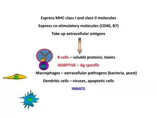
Antigen-presenting cells cells that can process and present antigens (MHC-peptide) to T cells Professional APC Dendritic cell Macrophage B lymphocyte nonprofessional APC • Several other cell types, classified as nonprofessional antigen-presenting cells, can be induced to express class II MHC molecules or a co-stimulatory signal Many of these cells function in antigen presentation only for short periods of time during a sustained inflammatory response.
APC • APC can express MHC-II and co-stimulatory molecules and present exogenous antigens to CD4+ T cells, besides presenting endogenous antigens to CD8+ T cells.Three cell types are classified as professional antigen-presenting cells: dendritic cells, macrophages, and B lymphocytes.
1. Dendritic cells Dendritic cells are bone marrow-derived cells Classification By source Myeloid DC Lymphoid DC By mature Immature DC Mature DC
1. Dendritic cells Dendritic cells are bone marrow-derived cells Classification By source Myeloid DC Lymphoid DC By mature Immature DC Mature DC By distribution • Lymphoid tissues Interdigitating DC, follicular DC • Non-lymphoid tissues Langerhans cell • Body fluid
滤泡树突细胞(follicular DC, FDC) FDC 淋巴滤泡内的FDC通过Fc受体和补体受体捕获被致敏的抗原,并将其递呈给B细胞 B cells
并指状树突细胞(interdigitating DC) IDC表达高水平的II类MHC分子和共刺激分子B7,具有激活T细胞的能力。
郎格汉斯细胞 (Langerhan’s cells) 上皮组织中的LC,捕捉外来抗原后即进入引流淋巴结的T细胞区,成为IDC
Function of DC : 1. Capturing and processing antigens 2. Presenting antigens During the maturation of DC , its ability of Ag capture and processing decreased while its ability of Ag presenting give a rise.
Function : 1. Phagocytosis 2. Presentation of antigens Nonactivated macrophage activated macrophage: MHC II molecules and costimulatory molecules
B cells Functions • Mediate humoral immune response • Present antigens to T cell Soluble Ag Specific receptor-mediated endocytosis
These cells differ from each other in their mechanisms of antigen uptake, in whether they constitutively express class II MHC molecules, and in their co-stimulatory activity: • Dendritic cells are the most effective of the antigen presenting cells,constitutively expressing class II MHC molecules and the costimulatory B7 molecule. • Macrophages must be activated by phagocytosis of • particulate antigens before they express class II MHC • molecules or the co-stimulatory B7 membrane molecule. • B cells constitutively express class II MHC molecules but must be activated before they express the co-stimulatory B7 molecule.
II. Processing and presentation pathway MHC class II pathway-------exogenous antigens MHC class I pathway-------endogenous antigens Cross-presentation pathway Non-classical pathway
MHC calss II pathway 1. Capture and processing of exogenous Ag 2. Synthesis and transportation of MHC II molecules 3. Formation of peptide - MHC II molecule complex 4.Presentation of peptide - MHC II molecule complex to CD4+ T cells
Capture and processing of exogenous Ag Exogenous antigens are endocytosed and the endosome is formed endocytosis: phagocytosis: particles or granules pinocytosis: liquids receptor-mediated endocytosis: specificexogenous antigen Endosome A vesicle is formed by partial cell membrane which surrounds the endocytosed antigens
EndosomeA vesicle is formed by partial cell membrane which surrounds the endocytosed antigensA phagolysome is formed when the endosome is fused with lysosome cathepsinsAg antigen peptidesThe antigen is hydrolysed into peptides by various proteases, such as cathepsins.
2. Synthesis and transportation of MHC II molecules Synthesis of MHC II molecules in ER Ii chain interacts with the pepetide-binding cleft of MHC II molecule ,Preventing any peptides from combining with MHC II molecules within ER Ii leads MHC II molecules into endosome from ER via Golgi complex Endosome (MIIC)
3. Formation of peptide - MHC II molecule complexThe Ii in the Ii-MHC II molecules complex is degraded in endosomeprotease Ii chain cleaving CLIP remains bound to the MHC class II molecules ( CLIP-MHC II molecules)HLA-DM catalyzes the exchange of CLIP with antigenic peptides. HLA-DM CLIP releasingAntigen peptide-MHC II molecules
4.Presentation of peptide - MHC II molecule complex to CD4+ T cellsantigen peptide-MHC II molecuels presented on cell membrane by exocytosis
MHC I pathway 1. Processing of endogenous Ag 2.transporting of antigen peptides into ER 3.Synthesis and assembly of MHC class I molecules 4. Formation and presentation of peptide- MHC molecules
1.Processing of endogenous Ag proteosome A multifunctional protease complex immunoproteosome A proteosome containing three subunits, PMSB-8,PMSB-9 and PMSB-10 preferentially generate peptides that bind to MHC class I molecules. Peptides that bind to MHCclass I molecules terminate almost exclusively with hydrophobic or basic residues.
2.transporting of antigen peptide into ERTAP(transporter associated with antigen precessing): Consisting of TAP1 and TAP2 ATP dependent transporter Selective transporting
3. Synthesis and assembly of MHC class I moleculeschaperone moleculescalnexin--- α chain of MHC moleculecalreticulintapasin---------------α and β2 microglobulin
4. Formation and presentation of peptide- MHC moleculesAs a consequence of peptide binding, the class I molecule displays increased stability and can dissoaciate from calreticulin and tapasin, exit from the ER and proceed to the cell surface via the Golgi.ERGolgi complex Exocytic vesicles Cell membrane
Ag(cytosolic protein) Proteasome proteolytic degradation Ag peptide TAP complex transporting into ER antigen peptide-MHC I molecule Golgi complex exocytosis Presenting to CD8+ T cells
Cross-presentation pathwy • In contrast to traditional presentation, exogenous antigens are presented by MHC class I pathway, or endogenous antigens are presented by MHC class II pathway.
Processing and presentation of antigens • I. APC (antigen presenting cells) • II. Processing and presenting pathway