Download
1 / 90
920 likes | 1.39k Vues
Bones & Joints of the Upper Limb. Kaan Yücel M.D.,Ph.D. 3 November 2011 Thursday. BONES OF UPPER LIMB. The pectoral girdle and bones of the free part of the upper limb form the superior appendicular skeleton.

E N D
Bones & Joints of the Upper Limb Kaan Yücel M.D.,Ph.D. 3 November 2011 Thursday
BONES OF UPPER LIMB • Thepectoral girdle and bones of the free part of the upper limb form the superior appendicular skeleton. • The pelvic girdle and bones of the free part of the lower limb form the inferior appendicular skeleton.
The superior appendicular skeleton articulates with the axial skeleton only at the sternoclavicular joint, allowing great mobility.
Connects the upper limb to the trunk. The shaft of the clavicle has a double curve in a horizontal plane. Clavicle (Collar bone)
Its medial half is convex anteriorly, and its sternal end is enlarged and triangular where it articulates with the manubrium of the sternum at the sternoclavicular (SC) joint. Its lateral half is concave anteriorly, and its acromial end is flat where it articulates with the acromion of the scapula at the acromioclavicular (AC) joint. Clavicle (Collar bone)
The clavicle • Serves as a moveable, rigid support from which the scapula and free limb are suspended, keeping them away from the trunk so that the limb has maximum freedom of motion. • Fixing the strut in position • Forms one of the bony boundaries of the cervico-axillary canal (passageway between the neck and the arm), affording protection to the neurovascular bundle supplying the upper limb. • Transmits shocks (traumatic impacts) from the upper limb to the axial skeleton.
The conoid tubercle, near the acromial end of the clavicle, gives attachment to the conoid ligament, suspends the remainder of the upper limb is passively from the clavicle.
Near the acromial end of the clavicle is the trapezoid line, to which the trapezoid ligament attaches. • The subclavian groove (groove for the subclavius) in the medial third of the shaft of the clavicle is the site of attachment of the subclavius muscle. More medially is the impression for the costoclavicular ligament, a rough, often depressed, oval area that gives attachment to the ligament binding the 1st rib to the clavicle, limiting elevation of the shoulder.
Scapula (Shoulder blade) A triangular flat bone Lies on the posterolateral aspect of the thorax, overlying the 2nd-7th ribs.
The convex posterior surface of the scapula is unevenly divided by a thick projecting ridge of bone, the spine of the scapula, into a small supraspinous fossa and a much larger infraspinous fossa.
The concave costal surface of most of the scapula forms a large subscapular fossa.
The spine continues laterally as the flat expanded acromion (G. akros, point), which forms the subcutaneous point of the shoulder and articulates with the acromial end of the clavicle.
The deltoid tubercle of the scapular spine is the prominence indicating the medial point of attachment of the deltoid. The spine and acromion serve as levers for the attached muscles, particularly the trapezius.
Superolaterally, the lateral surface of the scapula has a glenoid cavity(G. socket), which receives and articulates with the head of the humerus at the glenohumeral joint. The beak-like coracoid process (G. korakōdés, like a crow's beak) is superior to the glenoid cavity.
The scapula has medial, lateral, and superior borders and superior, lateral, and inferior angles. The glenoid cavity is the primary feature of the head.
The shallow constriction between the head and the body defines the neck of the scapula.
The largest bone in the upper limb Articulates with the scapula at the glenohumeral joint. Artciulates with the radius and ulna at the elbow joint. Humerus (Arm bone)
The proximal end of the humerus has ahead, surgicaland anatomical necks, and greater and lesser tubercles. The spherical head of the humerus articulates with the glenoid cavity of the scapula. The surgical neck of the humerus, a common site of fracture, is the narrow part distal to the head and tubercles.
The greater tubercle is at the lateral margin of the humerus, whereas the lesser tubercle projects anteriorly from the bone. The intertubercular (bicipital) groove separates the tubercles and provides protected passage for the slender tendon of the long head of the biceps muscle.
The shaft of the humerus has two prominent features: the deltoid tuberosity laterally, for attachment of the deltoid muscle The oblique radial groove (groove for radial nerve, spiral groove) posteriorly, in which the radial nerve and deep artery of the arm lie.
The inferior end of the humeral shaft widens as the sharp medial and lateral supraepicondylar(supracondylar) ridges form and then end distally in the especially prominent medial epicondyleand thelateral epicondyle, providing for muscle attachment.
The distal end of the humerus—including the trochlea; the capitulum; and the olecranon, coronoid, and radial fossae—makes up the condyle of the humerus. The condyle has two articular surfaces: Laterally Capitulum(L. little head) for articulation with the head of the radius Medially Trochlea (L. pulley) for articulation with the proximal end (trochlear notch) of the ulna
Anteriorly, the coronoid fossa receives the coronoid process of the ulna during full flexion of the elbow.
Posteriorly, the olecranon fossa accommodates the olecranon of the ulna during full extension of the elbow.
Superior to the capitulum anteriorly, a shallower radial fossa accommodates the edge of the head of the radius when the forearm is fully flexed.
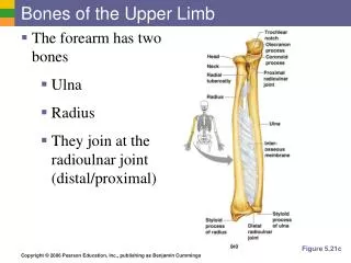
Bones of Forearm The two forearm bones serve together to form the second unit of an articulated mobile strut (the first unit being the humerus), with a mobile base formed by the shoulder, that positions the hand. However, because this unit is formed by two parallel bones, one of which (the radius) can pivot about the other (the ulna), supination and pronation are possible. This makes it possible to rotate the hand when the elbow is flexed.
Stabilizing bone of the forearm Medial and longer of the two forearm bones. Its more massive proximal end is specialized for articulation with the humerus proximally and the head of the radius laterally. ULNA
For articulation with the humerus, the ulna has two prominent projections: (1) olecranon, projects proximally from its posterior aspect (forming the point of the elbow). (2) coronoid process, projects anteriorly.
The olecranonand coronoid processesform the walls of the trochlear notch.
Inferior to the coronoid process is the tuberosity of the ulna for attachment of the tendon of the brachialis muscle. Inferior to the radial notch on the lateral surface of the ulnar shaft is a prominent ridge, the supinator crest. • On the lateral side of the coronoid process is a smooth, rounded concavity, the radial notch, which receives the broad periphery of the head of the radius.
Between the radial notch and the distal part of the coronoid process is a concavity, the supinator fossa.
At the distal end of the ulna a small, conical ulnar styloid process. • The ulna does not reach—and therefore does not participate • in—the wrist (radiocarpal) joint.
RADIUS • The radius is the lateral and shorter of the two forearm bones. • Its proximal end includes a short head, neck, and medially directed tuberosity.
Head of the radius articulation with capitulum of the humerus radial notch of the ulna
The neck of the radius is a constriction distal to the head. The oval radial tuberosity is distal to the medial part of the neck and demarcates the proximal end (head and neck) of the radius from the shaft.
The medial aspect of the distal end of the radius forms a concavity, the ulnar notch, which accommodates the head of the ulna.
The radial styloid process is larger than the ulnar styloid process and extends farther distally. • This relationship is of clinical importance when the ulna and/or the radius is fractured.
Bones of Hand The wrist, or carpus, is composed of eight carpal bones (carpals) arranged in proximal and distal rows of four. These small bones give flexibility to the wrist. The carpus is markedly convex from side to side posteriorly and concave anteriorly. Augmenting movement at the wrist joint, the two rows of carpals glide on each other; in addition, each bone glides on those adjacent to it.
From lateral to medial, proximal row of carpals • Scaphoid (G. skaphé, skiff, boat) • Lunate (L. luna, moon) • Triquetrum (L. triquetrus, three-cornered) • Pisiform (L. pisum, pea) • From lateral to medial, distal row of carpals • Trapezium (G. trapeze, table) • Trapezoid • Capitate (L. caput, head) • Hamate (L. hamulus, a little hook)
The proximal surfaces of the distal row of carpals articulate with the proximal row of carpals, and their distal surfaces articulate with the metacarpals.
The metacarpus forms the skeleton of the palm of the hand between the carpus and the phalanges. It is composed of five metacarpal bones (metacarpals). Each metacarpal consists of a base, shaft, and head.
Each digit has three phalanges except for the first (the thumb), which has only two; however, the phalanges of the first digit are stouter than those in the other fingers. Each phalanx has a base proximally, a shaft (body), and a head distally. The proximal phalanges are the largest, the middle ones are intermediate in size, and the distal ones are the smallest.
JOINTS OF UPPER LIMB • Movement of the pectoral girdle involves the sternoclavicular, acromioclavicular, and glenohumeral joints, usually all moving simultaneously. • Functional defects in any of the joints impair movements of the pectoral girdle.
Mobility of the scapula is essential for free movement of the upper limb. The clavicle forms a strut that holds the scapula, and hence the glenohumeral joint, away from the thorax so it can move freely.