Ch 8: The Cell Cycle
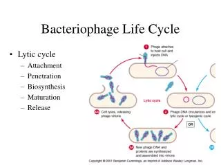
Ch 8: The Cell Cycle. From generation to generation. 1. Cell Division: The Cellular Copy Machine. 3. Did you know? Daughter cells are genetically identical to the parent cell!. Mitosis = Division of Nucleus Cytokinesis = Division of Cytoplasm. Binary Fission – Monerans 20.

Ch 8: The Cell Cycle
E N D
Presentation Transcript
Ch 8: The Cell Cycle From generation to generation... 1
Cell Division: The Cellular Copy Machine 3 Did you know? Daughter cells are genetically identical to the parent cell! Mitosis = Division of Nucleus Cytokinesis = Division of Cytoplasm
Binary Fission – Monerans20 Simple because there’s no….. …NUCLEUS!
The Cell Cycle: The life of a cell2 Mitosis and Cytokinesis Cycle Begins Gap 1 (Pre-replication): Cell grows and makes new proteins Gap 2 (Pre-mitosis): Cell prepares to divide Length varies widely See P. 215 What’s Interphase? G0: Non-dividing Cells (Eg. Neurons and adult cells) Restriction Point: “Point of No Return” Synthesis: Replication of DNA
DNA Structure I Phosphate group Nitrogenous base Sugar Nitrogenous base (A,G,C, or T) Nucleotide Thymine (T) Phosphategroup Sugar (deoxyribose) DNA nucleotide Polynucleotide Sugar-phosphate backbone 3 parts of a nucleotide Know which bases are 2 rings and which are 1 Figure 10.2
DNA Structure II • 3 parts of a nucleotide • Anti parallel structure • 5’ = Phosphate • 3’ = Sugar See P. 217
Chargaff’s Ratios 15 Edwin Chargaff – biochemist at Columbia Observed that in DNA, A % = T % and C % = G % So, scientists were able to figure out which bases were complementary… allowing decoding! 16
5 DNA in the Nucleus Liver Cell Nucleus –With visible chromatin 3 3 2 Condensed Chromosome During mitosis 1. Nucleosome = DNA + Histone Proteins 2. Chromatin (DNA Fiber) = Many nucleosomes 3. Chromosome = Highly folded chromatin 1 6 11_07AN Chromosome Structure.MOV
Origin of replication Origin of replication Origin of replication Parental strand Daughter strand Bubble Two daughter DNA molecules Semi-Conservative DNA Replication At the end of S Phase, all DNA has been duplicated! Figure 10.8 Each strand is ½ old and ½ new DNA
Steps of Synthesis 1. Forming the Replisome: Binding RNA Synthesis enzymes and DNA Polymerase enzymes to DNA at Replication Origin Unwinding (unzipping) DNA strand with Helicases 2. Stabilization with Single-Strand binding Protein DNA Synthesis: S Phase Prokaryotes use one origin; Eukaryotes use many at the same time! 4
4. Lagging Strand Synthesis Since DNA Pol can only work in one direction, Okazaki fragments are made on the lagging strand which are joined later by a ligase. 3. Leading Strand Synthesis DNA Polymerase needs a short strand of RNA (Primer) to get started… Then DNA Pol adds matching bases working in the 5’ to 3’ direction. 4 Adding new bases (correctly paired) and bonding them together.4 7 http://highered.mcgraw-hill.com/sites/0072437316/student_view0/chapter14/animations.html
DNA Repair – the never ending story Mutation = Change in sequence of DNA bases Mutagen = Something that causes mutation DNA Polymerase has 1 in 10,000 error rate DNA proofreading enzymes correct that to about 1 in 10 million 8 There are also other repair mechanisms that check the sequence before going on to mitosis!
Preparing for Mitosis After S Phase, the cell moves to G2 2 Copies of each chromosome (Sister Chromatids) are joined at the centromere Chromosome Segregation: Each chromatid will become part of a new nucleus during mitosis 4 Aneuploid Cells: Mistakes in mitosis make cells with abnormal # of chromosomes
Prophase4 • Nuclear envelope breaks down • Chromosomes condense • Microtubules form spindle • Centrioles (animal) or MTOC move to spindle poles, pushed by microtubules • Microtubules join kinetochore of each chromosome to centriole or MTOC 12SWF-CellCycle\12-05b-LateInterphase.swf 12SWF-CellCycle\12-05c-Prophase.swf
Chromosomes line up at metaphase plate (cell equator) perpendicular to spindle fibers Enzymes break down part of centromere Kinetochores pull chromatids along spindle towards poles Each separated chromatid is now called a chromosome Metaphase and Anaphase4 12SWF-CellCycle\12-05d-Prometaphase.swf 12SWF-CellCycle\12-05e-Metaphase.swf 12SWF-CellCycle\12-05f-Anaphase.swf
Telophase4 12SWF-CellCycle\12-05g-Telophase.swf
In Animals A Cleavage Furrow forms pinching one cell into two Cytokinesis begins in Anaphase In Plants Cell Plate begins to form making a new cell wall between the two cells Cytokinesis begins in telophase http://highered.mcgraw-hill.com/sites/0072437316/student_view0/chapter11/animations.html# Cytokinesis: Division of Cytoplasm 10 11 12 12SWF-CellCycle\12-05h-Cytokinesis.swf
Mitosis in Real Cells21 Scadoxus puniceus (Blood Lily, Snake Lily) 12SWF-CellCycle\08_07AN Mitosis Complete.MOV Mitosis Video Clips\Bloodlily.mov
Cell Fusion Experiments Something in the S/M cells causes the cell to advance from G0/G1 Cyclins: Proteins that accumulate and peak to tell cell to move on to next phase (See P. 226) Cell Cycle Control7
Kinases in the Cell Cycle Kinase = Enzyme that adds a Phosphate to a protein (Phosphorylation), changing its function 13 Look at P. 226 for G1 and M Cyclins Cyclins activate kinases --- Kinases phosphorylate proteins that • Function in cell cycle • Help produce enzymes/proteins needed in the cell cycle See P. 227
Checkpoints7 • Times where the cell looks at itself and chromosomes and stops if anything is wrong. • Problems are fixed before the cell continues on through the cycle. G1 Arrest: damaged DNA S Arrest: unreplicated DNA M Arrest: defective spindle G2 Arrest: damaged DNA
Sources Cited 1. http://www.micro.utexas.edu/courses/levin/bio304/genetics/celldiv.html 2. learninglab.co.uk/headstart/ cycle3.htm 3. www.denniskunkel.com with permission 4. http://www.emc.maricopa.edu/faculty/farabee/BIOBK/BioBookTOC.html 5. biologica.concord.org/.../ v2_top_right_chromatin_DNA.htm 6. http://imsb.au.dk/~raybrown/chromatin.gif 7. BioCD. From Biology, Fifth Edition. Campbell, Reece, Mitchell. Addison, Wesley, Longman. 1999. 8. http://www.ems-us.org/dnarep.jpeg 9. http://www.colorado.edu/epob/epob1030cornwall/fall_2001/figure%2009-08-1.gif 10. http://www.thomsonlearning.com.au/biology/guide/unit_4/unit1111.html?/biology/guide/unit_4/s1a.html~maintext 11. http://biog-101-104.bio.cornell.edu/BioG101_104/tutorials/cell_division/mito_q6_premise.html 12. http://www.biochem.wisc.edu/bednarek/ 13. http://www.blc.arizona.edu/marty/181/181Lectures/Figures/POHS_Figures/Chap9/phosphatase.gif 14. http://www.zo.utexas.edu/faculty/sjasper/images/john6.16.jpg 15. http://cpmcnet.columbia.edu/news/in-vivo/Vol2_Iss10_may26_03/CPMC-history.html 16. www.yourgenome.org/ intermediate/all/ 17. http://web.mit.edu/doroquez/www/7.014/archive.html 18. BSCS Biology Blue, 8th Edition 19. http://www.bio.miami.edu/dana/250/25003_20.html 20. Bioshow: for Biology: Concepts and Connections, Second Edition. Campbell, Mitchell, and Reece 21. http://www.museums.org.za/bio/plants/amaryllidaceae/scadoxus.htm