Download

1 / 68
720 likes | 1.22k Vues
Eukaryotic Chromosome Organization — Lecture II. Dr. Steven J. Pittler VH 375B Office 4-6744 Cell 612-9720. Suggested Reading: Lewis 2nd Edition Chapter on Chromosomes. Chromosomes.

E N D
Eukaryotic ChromosomeOrganization — Lecture II • Dr. Steven J. Pittler • VH 375B • Office 4-6744 • Cell 612-9720 Suggested Reading: Lewis 2nd Edition Chapter on Chromosomes
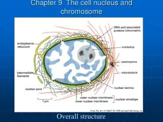
Chromosomes • Structures that consists primarily of DNA and proteins that are duplicated and transmitted during mitosis or meiosis • Heterochromatin stains dark and is mostly repetitive DNA sequences • Euchromatin stains lighter and contains protein encoding genes
Multiple Levels of packing are required to fit the DNA into the cell nucleus
The basic unit of chromatin is the nucleosome
The nucleosome consists of 146bp of DNA wrapped around a protein core of 8 histones
Histone H1 helps compact the nucleosomes into a 30nm fiber
The Histone tails are a critical determinant of chromatin structure
Specific modifications are associated with specific functions
The normal karyotype: 23 diploid chromosomes • Human somatic cells contain 46 chromosomes: • paired homologs of chromosomes 1 to 22 and • sex chromosomes (XX or XY) • • Diploid refers to the presence of two copies of each different chromosome. • Gametes have one set of each chromosome and are called haploid. • Cells missing a single chromosome or having an extra one are aneuploid • Cells which contain a normal chromosome constitution are called euploid.
Karyotype analysisMetaphase chromosomes are squashed on a slide and stained with DNA binding dyes. Banding patterns help define different chromosomes. Chromosomes were named in order of their size and centromere position that appear during mitotic metaphase.
“Histone Code” hypothesis Modifications of the Histone tails act as marks that can be read by other proteins to control the expression or replication of chromosomal regions. The coding in the histones may be heritable. E.g. Generally, histone acetylation is associated with transcriptionally active genes Deactylation is associated with inactive genes (= gene silencing)
Epigenetics Heritable changes not caused by mutation in the DNA Can be due to stable changes in gene expression caused by changes in chromatin structure
Epigenetics and Disease: Genomic imprinting Parent specific expression or repression of genes or chromosomes in offspring. So… even though two copies of a given gene are inherited, one from each parent, only the maternal or paternal allele is expressed. The non-expressed allele is said to be “imprinted.”
Wilms Tumor • -Childhood Tumor of the kidney (nephroblastoma) • Accounts for 7% of all childhood cancers • -Caused by a defect in imprinting of the Insulin-like • Growth Factor 2 (IGF2) gene • IGF2 is usually only expressed from the paternal locus, i.e. • maternally imprinted • -Defects in imprinting that cause expression of the maternal • locus lead to cancer
DNA methylation Covalent modification of the DNA is also important for gene silencing human cells Most genes have GC rich areas of DNA in their promoter regions. These are referred to as CpG islands. Methylation of the C residues within the CpG islands leads to gene silencing
Epigenetics and Disease: DNA methylation Rett Syndrome: X-linked, neurodegenerative disorder affects 1:10,000-15,000 (females only) Caused by a mutation in the gene encoding Methyl-CpG-binding protein 2 (MeCP2), which in turn leads to loss of gene silencing at many loci.
The karyotype shows the chromosome complement of a normal ________? • Male • Female
Largest, Metacentric Smallest acrocentric Sex chromosomes XX (shown)
Anatomy of a chromosome • Chromosomes are categorized by the relative location of their centromere. • At tip - telocentric (not found in humans) • Close to tip - acrocentric • At midpoint - metacentric • Displaced from center - submetacentric
Anatomy of a chromosome • The portion of the chromosome to each side of a centromere is called a chromosome arm. shorter arm = p arm longer arm = q arm
Anatomy of a chromosome • Telomeres are: • At the tips of chromosomes • Many repeats of the sequence TTAGGG • Subtelomeres have more varied short repeats • These are the chromosomal parts between protein-rich areas and the telomeres • These areas extend from 8,000 to 30,000 bases inward toward the centromere from the telomeres
Subtelomeres • Include some protein-encoding genes and bridge the gene-rich regions and the telomere repeats • When researchers compared subtelomeres to known gene sequences they found about 500 matches
Anatomy of a chromosome • Centromeres are the largest constriction of the chromosome • Site of attachment of spindle fibers • 171 base pair segment repeated 100,000 times, called alpha satellite sequences • Also include centromere-associated proteins • Some are synthesized only when mitosis is imminent forming the kinetochore that emmanates from the centromere and connects the spindle fibers • Appears during prophase and vanishes during telophase
Chromosomal shorthand • An ideogram represents a chromosome schematically. • The major banding regions are indicated with numbers. • Sucrose intolerance is located at 3q.26 • (chromosome 3, long arm, major band 26)
Chromosomes carry different genes ideograms
Visualizing chromosomes • Obtain tissue from person • Fetal tissue: amniocentesis • chorionic villi sampling • fetal cell sorting • Adult tissue: blood (white blood cells) • cheek swab (buccal cells) • skin cells • tissue biopsy • Prepare cells on slide to remove rest of cell matter • Stain DNA with dyes or DNA probes (is a labeled piece of DNA that binds to its complimentary sequence on a particular chromosome) to visualize DNA • Evaluate chromosomes in comparison to known information
FISHing • Conventional chromosome stains have one drawback- they are not specific to a particular chromosome • FISH uses DNA probes that are complimentary to specific base sequences, and if those sequences are unique to a particular chromosome the technique can identify it
FISH: fluorescence in situ hybridizationDNA probes labelled with fluorescing dye bind complementary DNA
Cytogenetics • the subdiscipline within genetics • that focuses on chromosome variations. Abnormal number of copies of genes or chromosomes can lead to genetic abnormalities.
Mutation at the Chromosome Level • Abnormal numbers of genes or chromosomes • Range from the single-base changes to missing or extra pieces of chromosomes • A mutation is a chromosomal aberration if it is large enough to see with a light microscope using stains and/or fluorescent tags • Generally, excess genetic material has a milder effect on human health when compared to a deficit of genetic material • Most chromosomal abnormalities are so severe that prenatal development ceases in the embryo • 0.65 percent of all newborns have chromosomal aberrations • An additional 0.20 percent have chromosomal rearrangements that do not produce symptoms
Chromosomal Abnormalities • Down syndrome-extra chromosome 21 • Turner syndrome- XO- a female with only one X chromosome • Klinefelter syndrome- XXY, a male with an extra X chromosome • Before this women who were XO were thought to be genetic males because they lack Barr bodies and XXY were thought to be genetic females because their cells have Barr bodies
Chromosome anomaliesmay cause phenotype abnormalities. • A chromosome karyotype revealed she carries three copies of chromosome 21, a condition called trisomy 21. This young girl has Down syndrome.
Extra Autosomes • Chromosomes 13, 18, and 21 are the most frequently seen extra autosomes and they have the lowest gene densities- they carry considerably fewer protein-encoding genes
Polyploidy Aneuploidy monosomy trisomy Deletion Duplication Inversion Translocation Iso chromosome Ring chromosome Extra chromosome set Extra or missing chromosome one chromosome absent one chromosome extra Part of a chromosome missing Part of a chromosome present twice Segment of chromosome reversed Two chromosome arms exchanged in part or entirely A chromosome with identical arms A chromosome that forms a ring due to deletions in telomeres, which cause ends to adhere Chromosome Abnormalities
Polyploidy • Individuals with three copies of each chromosome are triploid, or an extra set • Polyploidy accounts for 17% of all spontaneous abortions and 3% of stillbirths/newborn deaths. • Result of: • Two sperm fertilize one egg. • Haploid sperm fertilizes diploid egg.
Aneuploidy • Most autosomal aneuploids are spontaneously aborted • Mental retardation is common in an individual who survives aneuploidy • Sex chromosome aneuploidy have milder symptoms • Children born with the wrong number of chromosomes have an extra chromosome- trisomy • Rather than missing a chromosome- monosomy • Down syndrome can result from trisomy 21 or from translocation • Translocation Down syndrome accounts for 4% of cases, has a much higher risk of recurrence than trisomy 21
Aneuploidy • Nondisjunction is a common cause of aneuploidy resulting in a gamete with one extra chromosome and another gamete with one missing chromosome. • Nondisjunction during the first meiotic anaphase division results in a copy of each homolog in the gamete and two cells do not have any copies. • Nondisjunction during the second meiotic anaphase division results in both sister chromatids in one gamete, one with no copy, and two normal cells.
Abnormal gametes Abnormal gametes Normal gametes Nondisjunction causes aneuploidy Nondisjunction in meiosis I Nondisjunction in meiosis I Anaphase I Nondisjunction in meiosis II Anaphase II Gametes
Trisomies and Monosomies • One extra or one missing chromosome results in extra or missing copies of all of the genes on that chromosome. • Most trisomies and monosomies produce inviable embryos. • Some fetuses with trisomy of smaller autosomes survive to birth with syndromic conditions:
Autosomal Aneuploids • Most autosomal aneuploids are lethal • Trisomy 21 Down syndrome • Most common • Extra folds in the eyelids called epicanthal folds and a flat face • Termed mongoloid by Sir John Langdon Haydon in 1866 • In 1961, researchers identified a mosaic Down syndrome • Affected girl with all the physical signs but normal intelligence
Autosomal Aneuploids • Trisomy 21 cont’d • Usually short and has straight, sparse hair, and a thick tongue protruding through the lips • Hands have abnormal pattern of creases, loose joints, and poor reflexes and muscle tone give a floppy appearance • Intelligence varies • Physical problems are common • Heart and kidney defects, and hearing and vision loss • Suppressed immune system • Digestive system problems • Down syndrome 15 is more likely to develop leukemia
Autosomal Aneuploids • Trisomy 18- Edward Syndrome • Only 1 in 6,000 -10,000 newborns have trisomy 18 • Most do not survive birth • Great physical and mental disabilities, with developmental skills stalled at the six-month level • Major abnormalities • Heart defects, displaced liver, growth retardation, and oddly clenched fists • Overlapping placement of fingers, narrow and flat skull, abnormally shaped and low-set ears, small mouth and face, unusual or absent fingerprints, short large toes with fused second or third toes, and “rocker-bottom” feet • Most cases are attributed to non-disjunction in meiosis II of the oocyte
Autosomal Aneuploids • Trisomy 13- Patau Syndrome • Most do not survive birth • Most striking but quite rare is fusion of the developing eyes, so that the fetus has one large eyelike structure in the center of the face • More common is a small or absent eye • Major abnormalities • Heart defects, kidneys, brain, face, and limbs • The nose is malformed, and cleft lip and/or present in a small head • Extra fingers and toes • Extra spleen, abnormal liver, rotated intestines, and an abnormal pancreas
Turner syndrome • 45, X • 1 in 2,000 female births • 99% of Turner die in utero • Absence of Y leads to development as a female. • Absence of two copies of X-linked genes in a female results in Turner syndrome. • Phenotypes include short stature, webbing at back of neck, incomplete sexual development, hearing impairment, malformed eyebrows.
Turner syndrome • A chromosomal imbalance causes the hormone deficit • 1954 P.E. Polani discovered cells from Turner syndrome patients lack a Barr body (inactive X) • 50% are XO, the rest have partial deletions or are mosaics, with only some cells affected • Like autosomal aneuploidy this syndrome is more frequent among spontaneously aborted fetus than newborns • Two X chromosomes are necessary for normal sexual development