Major Pelvic Trauma
300 likes | 620 Vues
Major Pelvic Trauma. Bernard Foley FACEM Department of Emergency Medicine Auckland Hospital Friday, 3 October 2014. The Issues. Pelvic trauma doesn’t come in on it’s own Routine Pelvic x-ray in blunt trauma Do we always need it? The unstable patient Fracture instability

Major Pelvic Trauma
E N D
Presentation Transcript
Major Pelvic Trauma Bernard Foley FACEM Department of Emergency Medicine Auckland Hospital Friday, 3 October 2014
The Issues • Pelvic trauma doesn’t come in on it’s own • Routine Pelvic x-ray in blunt trauma • Do we always need it? • The unstable patient • Fracture instability • Haemodynamic instability • Prioritising interventions • No universal algorithm
Anatomy Sacrospinous ligament Sacrotuberous ligament SI joint and ligaments Pubic symphisis
Pelvic Fracture Types Lateral Compression B2 type partially stable Vertical Shear C1 type unstable AP Compression B1 type partially stable
Haemodynamic stability is the key • Unstable • Definitive haemostatic procedure • Assisted stability • Investigations to target interventions • Stable • Investigation cascade
Sources of bleeding in pelvic trauma • Arterial • Usually laceration/avulsion associated with ligamentous injuries • Mx therapeutic embolisation • Venous • Mx orthopaedic • Osseous • Mx orthopaedic
Anterior division branches of internal iliac most commonly injured Internal pudendal : between SSL and STL Inferior gluteal : above SSL Obturator : through foramen Posterior division branches of internal iliac artery most commonly injured Superior gluteal : piriformis fascia or sacral # Ilio-lumbar : sacral/ SI joint injuries Sources of arterial bleeding in pelvic trauma
Orthopaedic trauma Auckland Hospital 1995-2000 • 6040 orthopaedic trauma admissions • 520 Pelvic fractures • 45% transfers
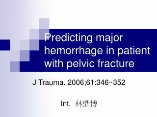
Pelvic trauma in Auckland hospital • 1 Jan 1995-31 Dec 1998 • 364 pelvic fractures • 76 Haemodynamically unstable • Mean ISS 30 (9-66) • 39/76 car crash • 10/76 motorcycle • 8/76 pedestrian • 13/76 falls • 27/76 deaths
Injury patterns • 43.7% Type A • 28.5% Type B • 27.8% Type C • 49 Mechanically unstable pelvic injuries / year
Associated injuries • Chest / abdomen 23% • Genitourinary 17% • Head injury 31%
Associated injuries • Sacral nerve injuries • Rectal perforation • Vaginal perforation • Bladder and vesical injuries • Spinal injuries • Femoral fractures • Long-term disability
Mortality • Uncontrolled haemorrhage • Chest • Abdomen • Retroperitoneal • Other unsurvivable injuries • i.e. neurological injury • Multiorgan failure • Sepsis
Multitrauma / Time critical • Structured approach required • A,B,C’s • Resuscitation • Trauma radiography • Hx, examination, Ix • Extended trauma team concept • Interventional radiology • Orthopaedics • Urology
Pelvic trauma x-ray • Currently recommended as part of trauma series • Gonzalez et al (n=2,176) • Alert patients (GCS14-15), blunt trauma • Ethanol levels 16-75mmol/L (n=463) • 97 patients with pelvic fractures • Physical exam sensitivity 93% • No significant fractures missed • Pelvic x-ray sensitivity 87% • 6 requiring operative intervention • J Am College Surg 194,No2. Feb 2002
CT scanning • Good at assessing haemorrhage in peritoneum and retro peritoneum • Can aid planning of vascular/orthopaedic procedures • Good at assessing pelvic fractures • Requires stable patient (?assisted stability)
Procedures-pelvic • Sheet wrap • External fixation • Internal fixation • Angiography
Quick and easy Inexpensive Can do in ED Good tamponade of expanding haematoma Not definitive stabilisation May impact on exposure Sheet wrap
External fixation • Good control of anterior instability • Dependent on bone quality • Not definitive • Impairs mobilisation • Can burn some bridges
Open internal fixation • Big exposures • Unavoidable complication rate • Timing problematic in multitrauma
Exposure not a problem Low complication rate Bio mechanically ideal Detailed anatomical knowledge required Technically demanding Percutaneous fixation
Therapeutic embolisation • Selective IIA angiography shows higher incidence and severity of bleeding than aortic flush studies • Better pickup of hypo-perfusion and spasm
Method of EmbolisationAnterior Division Embolisation • Proximal embolisation more effective • Adverse events rare • Buttock claudication
Therapeutic embolisation • Allows ancillary procedures • i.e. percutaneous nephrostomy
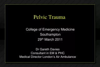
Pelvic Fracture: Patient Haemodynamically unstable no no yes yes yes yes no no
Summary 1 • A-P pelvis radiograph • GCS <14 • Clear clinical evidence of fracture • Suspicious mechanism • ? Validated set of rules
Summary 2 • Early involvement of orthopaedic and Interventional radiology • Prioritisation of interventions • Early haemodynamic instability= arterial bleeding= interventional radiology • Assisted stability may buy time for additional investigations • Early percutaneous fixation appears to produce the best results