Download
1 / 25
250 likes | 282 Vues
This lecture explores the basics of bioinformatics databases and tools, including sequence searches, multiple alignments, and phylogenetic trees. It also focuses on the importance of protein domains and 3D structure in understanding protein function.

E N D
Bioinformatics for biomedicineProtein domains and 3D structure Lecture 4, 2006-10-10 Per Kraulis http://biomedicum.ut.ee/~kraulis
What is bioinformatics? Basic databases and tools Sequence searches: BLAST, FASTA Multiple alignments, phylogenetic trees Protein domains and 3D structure Seminar: Sequence analysis of a favourite gene Gene expression data, methods of analysis Gene and protein annotation, Gene Ontology, pathways Seminar: Further analysis of a favourite gene Course design
Multiple alignment • Example: ras proteins • FASTA input file available at course site • http://www.ebi.ac.uk/Tools/sequence.html • ClustalW, MUSCLE • http://msa.cgb.ki.se/cgi-bin/msa.cgi • Kalign
Proteins and drug action • Most drugs act via proteins • Binds to the protein • Inhibits or activates it • “Drug targets” • Small subset of all proteins • Certain protein families • 3D structural basis
3D structure and protein function • ‘Native’ state • Functional state • Well-defined 3D structure (usually) • Folded state • Denaturing a protein inactivates it • Heat, salt, solvents… (boiling an egg) • Undefined 3D structure; a mess • Unfolded state
3D structures are complicated • 1000’s of atoms • Hard to see anything • But: details essential for understanding
Levels of 3D structure • Primary • Protein sequence • Secondary • Alfa helix, beta sheet; hydrogen bonds • Tertiary • Overall 3D structure; “fold” • Quaternary • Complex between biomolecules
Simplified view of 3D structure • Schematic view • Peptide chain • Secondary structure • Alfa helix • Beta strands • Ligands • Metal ions • Cofactors • Drugs
Structure database • PDB database • 3D coordinates for structures • Protein, DNA, RNA, complexes • Experimentally determined structures • At RCSB: http://www.rcsb.org/pdb/ • Via UniProt: http://www.ebi.uniprot.org/ • Many different 3D viewers…
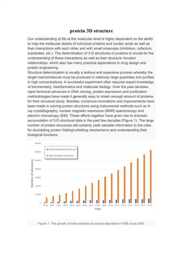
Experimental determination • X-ray crystallography • Atomic resolution • Small and large molecules (ribosome) • NMR • Small, soluble proteins • Electron microscopy • Not atomic resolution • Very large molecules/particles
X-ray crystallography 1 • Requires protein crystal • Size 0.1-1.0 mm • Possibly with ligands, other proteins • Optimize conditions (salt, solvents,…) • Recent technological advances • Structural Genomics Initiatives
X-ray crystallography 2 • Quality? • Resolution • How much data was available? • Given in Ångström • Good: 2.0 Å or less • Bad: 3 Å or higher • Refinement • How well do atomic coordinates agree with data? • R-value • Should be 20% or less
X-ray crystallography 3 • Is crystal state relevant? • Short answer: Yes • High solvent content; similar to cell plasma • Comparisons indicate OK • But: occasional relevant differences • Surface differences; loops • Details of ligand binding
Protein structure and prediction • Secondary structure prediction • Typically 70-80% accurate • Not as good as it sounds • Always use experimentally derived data, when available • 3D structure modelling • Based on known structure: predict similar • OK when sequence >90 % similar • Depends, when 50-90% similar • Problematic, when <50% similar
The folding problem 1 • 3D structure encoded in protein sequence • Anfinsen 1960s • Small proteins refold spontaneously • Synthetic peptides fold • Activity as for ‘natural’ protein • Assisted folding does not change this • Chaperones: catalysts for folding • Needed in cell environment (thick soup)
The folding problem 2 • Protein sequence determines 3D structure • 3D structure should be predictable • But: notoriously hard (unsolved) problem Physics Native protein Denatured protein Prediction 3D structure Protein sequence
Protein domains 1 • Proteins appear to be modular • 3D structure: pearls on a string • Sequence: partial sequence similarity
Protein domains 2 • Proteins are modular • Particularly in eukaryotes • A part of sequence as a unit • The units are called domains • Partial gene duplication • Domain families • Proteins containing a particular domain
Protein domains 3 • Sequence-based domain definitions • Pfam http://www.sanger.ac.uk/Software/Pfam/ • Based on Hidden Markov Models (HMMs) • Statistical method to detect patterns • Sensitive • Other domain databases • Structural: CATH http://www.cathdb.info/
Protein domains 4 • Domains from structure or sequence? • Usually very similar results • But some differences • Sequence region inserted • Structure formed from different parts of sequence
Protein families in medicine • “Druggable” proteins • Contains domains known to bind drugs • Small subset of proteins • Usually: binds small molecules naturally • Enzymes • Receptors • Protein-protein interactions very difficult • Much work, few results
Example: Nuclear receptors • Transcription factors • 3D structures available for several • Small-molecule ligands • Estrogen, progesteron,… • Androgen receptor • ANDR_HUMAN, P10275 • Several drugs • Tamoxifen
Example: 7TM receptors • 7TM = Seven transmembrane helices • Receptors • CNS, other systems • Maybe 50% of drugs act on these • Integral membrane protein • Very hard for X-ray crystallography • No good 3D structure!
7TM: rhodopsin • Light detector protein in eye • 3D structure determined! • Serves as model for other 7TM proteins • Known to be very approximate • UniProt: OPSD_BOVIN, P02699 • Retinal ligand shows binding pocket • In some 7TM proteins: different binding site
Example: protein kinases • Regulatory proteins, signalling • Cancer • Much recent work • Few drugs on market, yet • Drug design strategy • Compete with ATP in its pocket • Specificity? • UniProt: ABL1_HUMAN,P00519 • PDB: 2F4J