Download
1 / 73
730 likes | 761 Vues
Explore recombinant DNA techniques such as creating cDNA, using restriction sites, and genomic library construction. Learn about Southern/Northern blot analysis, sequencing, and genetic engineering applications. Discover how to detect specific genes in libraries and create transgenic organisms.

E N D
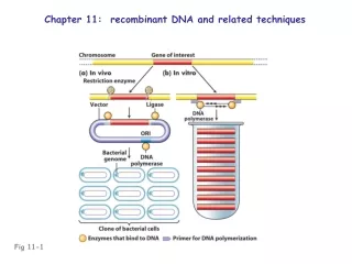
Recombinant (chimeric) DNA: fused DNA from two different organisms Recombinant clone: vector (bacterial plasmid, virus) + insert (DNA fragment to be cloned) Recombinant (transgenic) organisms: host genome + clone from another organism
cDNA: “complementary DNA”; DNA • complementary to RNA • Usually made against mRNA • cDNA is essentially an intron-less copy of a gene, • minus 5’ and 3’ flanking regulatory regions of the • gene • Prepared using reverse transcriptase (an RNA- • dependent DNA polymerase enzyme of RNA viruses)
Creating cDNA (DNA complementary to mRNA) Fig 11-2
Creating cDNA (DNA complementary to mRNA) Fig 11-2
Creating cDNA (DNA complementary to mRNA) Fig 11-2
Creating cDNA (DNA complementary to mRNA) Creates clonable DNA copy of specific mRNA or can make cDNA library (representing mRNA population) Fig 11-2
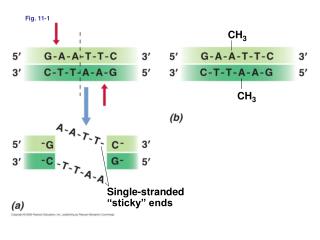
Using restriction sites to create a recombinant molecule Fig 11-3
4-6 4-4 pallindromic sequence cohesive ends Fig 11-4
Using restriction sites to create a recombinant molecule Fig 11-5
Using restriction sites to create a recombinant molecule Fig 11-5
Cells receiving a complete plasmid form colony Grow and purify DNA from single colony Fig 11-6 Useful for inserts <10kb
Using antibiotic resistance markers to select plasmid-bearing colonies Fig 11-6
Bacteriophage lambda: engineered as vector • for cloning large DNA fragments • Central 1/3 of genome (~45 kb) contains • lysogenic function genes • Can substitute ~15 kb cloned DNA into • genome and the virus is still capable of • lytic infection • e.g., the Drosophila genome (~150,000 kb) can be contained in • a minimum of 10,000 recombinant lambda clones (can fit on one • 15 cm Petri plate)
Creating a genomic library in bacteriophage lambda Useful for inserts 10-20kb Fig 11-7
Fig 11-8 Useful for inserts 100-300kb
Identifying a desired clone/gene in a library: • Use a probe (previously cloned DNA, oligonucleotide, • or antibody)
Detecting & isolating a specific clone within a library by hybridization Fig 11-11
Using an antibody to detect & isolate a specific clone within a library Fig 11-1
Identifying a desired clone/gene in a library: • Use a probe (previously cloned DNA, oligonucleotide, • or antibody) • Functional complementation (useful in organisms • with small genomes) • Positional cloning (chromosome “walk” to mutant • rearrangement site)
Chromosome walking to identify/isolate a region containing a gene Fig 11-15
Agarose gel electrophoresis • separates DNA fragments • by size: • restrict cloned DNA • electrophoresis • stain with ethidium bromide • visualize under UV Fig 11-13
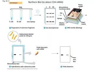
Southern/Northern blot analysis • agarose gel electrophoresis • transfer to nitrocellulose • hybridize with radioactive probe • autoradiograph to • detect bands containing • probe sequence Fig 11-14
Using restriction sites as markers to map a DNA fragment Fig 11-16
Using restriction sites as markers to map a DNA fragment Fig 11-16
Sanger dideoxy DNA sequencing Fig 11-18
Sanger dideoxy DNA sequencing • Mixture of ddATP + dATP • permits formation of • chains of various lengths • common 5’ end (primer) • vary by 3’ ends, marking locations of • A residues (T residues on template) Fig 11-18
Sanger dideoxy DNA sequencing Fig 11-18
Sanger dideoxy DNA sequencing Fig 11-18
Automated sequencing readout of Sanger dideoxy DNA sequencing Fig 11-19
An initial bioinformatic analysis Scan sequence for exceptionally long ORFs Fig 11-20
Polymerase chain reaction (PCR) • Uses heat-stable DNA polymerase • (e.g., Taq polymerase) • Requires two opposite-strand primers; • ~100 bp - ~3 kb apart on the target template • Uses a regimen of temperature cycling to amplify • the DNA target between the two primers
Polymerase chain reaction Specific primers permit specific amplification of a DNA segment Fig 11-21
Understanding alkaptonuria Fig 11-22
Detecting sickle-cell β–globin allele Fig 11-24
Detecting sickle-cell β–globin allele Heterozygote? Fig 11-24
Ti plasmid: a vehicle for making transgenic plants Fig 11-28
Inherited as a Mendelian dominant marker Fig 11-31
Engineering of mammalian genomes Insert a gene (relatively easy) Destroy a gene (“knockout”) Replace a gene (e.g., gene therapy)
Ectopic transformation of mouse embryos Insertions at random (ectopic) sites Fig 11-34
Making a targeted mutation (“knockout”) in mouse cells Fig 11-35
Making a targeted mutation (“knockout”) in mouse cells Fig 11-35
Making a targeted mutation (“knockout”) in mouse cells Fig 11-35