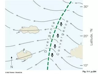
Fig. 11-1
130 likes | 308 Vues
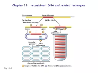
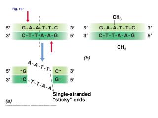
Fig. 11-1. CH 3. CH 3. Single-stranded “sticky” ends. Table 11-1. Fig. 11-2. Foreign DNA. Fig. 11-5. Cut with restriction enzyme. Add vector cut with same restriction enzyme. Sticky ends. Vector. Add DNA ligase to form recombinant molecules. Cloned DNA. Introduction

Fig. 11-1
E N D
Presentation Transcript
Fig. 11-1 CH3 CH3 Single-stranded “sticky” ends
Foreign DNA Fig. 11-5 Cut with restriction enzyme Add vector cut with same restriction enzyme Sticky ends Vector Add DNA ligase to form recombinant molecules Cloned DNA Introduction of recombinant vector into a host
Order of restriction enzyme cut sites in polylinker Ampicillin resistance Apo I - EcoR I lacZ Ban II - Sac I Fig. 11-6 Acc65 I - Kpn I Ava I - BsoB I - Sma I - Xma I BamH I Xba I Acc I - Hinc II - Sal I Polylinker BspM I - BfuA I Sbf I Pst I pUC19 2686 base pairs Sph I Hind III lacl Origin of DNA replication
Recombinant DNA = plasmid + insert • How do you know if insert is present? • Restriction digest • Blue/White selection
lacZ AmpR Fig. 11-7 Foreign DNA Vector Digestion with restriction enzyme Join with DNA ligase Opened vector Vector plus insert Recyclized vector without insert Transform into Escherichia coli and select on ampicillin plates containing Xgal Transformants blue (-galactosidase active) Transformants white (-galactosidase inactive)
Fig. 11-8 Missing OH Normal deoxynucleotide Dideoxy analog DNA chain Direction of chain growth No free 3-OH, replication will stop at this point
DNA strand to be sequenced Add DNA polymerase, mixture of all four deoxyribonucleotide triphosphates; separate into four reaction tubes Radioactive DNA primer Fig. 11-9 A small amount of only one dideoxynucleotide triphosphate (ddGTP, ddATP, ddTTP, or ddCTP) added to each tube and reaction allowed to proceed Reaction products ddGTP ddTTP ddATP ddCTP G A T C 7 6 Reaction products separated by electrophoresis on gel and identified by autoradiography 5 4 3 2 1 Sequence reads from bottom of gel as A G C T A A G. Sequence of unknown is 3 T C G A T T C 5 G A A A G C T
Copies of target gene(s) PCR cycle Target gene(s) 1 0 Fig. 11-11 DNA polymerase Heat Primers Primer extension 1 2 108 107 106 105 Copies of target gene 104 103 102 Repeat cycle 2 4 10 2 4 6 8 10 14 16 20 12 18 Repeat cycle 8 3 Number of PCR cycles
Copies of target gene(s) PCR cycle Target gene(s) 1 0 Fig. 11-11abc DNA polymerase Heat Primers Primer extension 1 2 Repeat cycle 2 4 3 8 Repeat cycle