Development
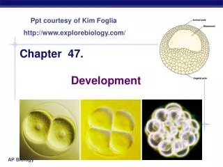
Development. Chapter 21, 46, 47 Mostly Human Development See pages 636 and 640 for overview of Phylogenetic tree. Early Development. Early Development: Developmental Tissue Layers, Sponges-0, Gridera /Centophores-2, All other Phyla-3 Hormones released in hypothalamus

Development
E N D
Presentation Transcript
Development Chapter 21, 46, 47 Mostly Human Development See pages 636 and 640 for overview of Phylogenetic tree
Early Development • Early Development: Developmental Tissue Layers, Sponges-0, Gridera/Centophores-2, All other Phyla-3 • Hormones released in hypothalamus • FSH: Follicle Stimulating Hormone • LH: Luteinizing Hormone • At menstruation, there are multiple concurrent events • Estrogen and Progesterone (hormones from the ovaries) gone down its concentration • FSH increases in concentration • Menstruation is occurring. The endometrial lining of the uterus is flushed out. • When Estrogen levels drop, the brain monitors and produces FSH. The target of the increased at FSH is in the ovaries. • A cluster of cells in the back of the ovary. Each contains the germ line cell. Primary oocye that started meiosis before birth. But this was arrested in prophase. • After 2 or 3 days, usually only one Oocyte continues to mature
The maturing of Oocyte 1. The surrounding cells form the GraffarianFollicle. 2. Continues into the first division of meiosis 3. the developing follicle moves down the Ovary and growing in cell number 4. the follicle continues to mature for about 14 days – it enlarges spatially and more cells • the meiotic dividing primary oocyte is in a cavity with some surrounding nursing cells. • The nursing cells are adding nutrients and other components to this meteoric cell to prepare for it to become an egg • fully developed follicle (egg/ovary) • outer follicle cells synthesis and release Estrogen (steroid) Maturing outer follicle becomes a hormone producer.. 5. Increased Estrogen stimulates the rebuilding of the Endometrium of the uteral wall and the decline of FSH concentration. 6. The brain monitors increased estrogen and as a feedback mechanism, it decreases the release of FSH.
The maturing of Oocyte 7. The developing follicle also can secrete inhibits, this is also a hormonal message to the hypothalamus and anterior pituitary to decrease the release of FSH. • An egg is being formed – both still requires 2nd meiotic division • the Estrogens and Inhibins decrease with equals FSH increases • Endometrium is rebuilding. The endometrium is continuing to form under hormonal direction and or stimulates currently estrogens. Mostly made of mucus, blood, glycogen. It's being formed in case of the pregnancy. If there is a pregnancy endometrium is the nutrient, food, 02 etc. source and will maintain the first part of the pregnancy. • This is for internal fertilization only not for things like fish as the ovarian follicle nears maturity, it's: • estrogenic output decreases • This is a signal to the brain or hypothalamus to cause anterior pituitary • release more FSH • release a very large amount of luteinizing hormone. • The estrogen decrease is not enough of drop to stop the endometrium from building • spike of LH stimulates to events to occur and a concurrent third event
Ovulation • Ovulation: Oocyte sent to the filopian tubes and ovaduct with nursing cells (triggers 2nd meiotic division) • Remaining follicle cells collapse in and forms the Corpus Luteum. • Two possible concurrent events during ovulation • the released egg travels down the ovaduct by Cilia on ovaduct cells and contraction of smooth muscle cells, called peristolsis. • The Corpus Luteum becomes the new Hormone Factories: prodcues Estrogen and Progesterone. - causes LH and FSH to decrease, causes Endometrium to enlarge more • (possibility) the egg can be fertilized, but the egg is viable (able to function) for about 24 hours • to become fertilized • sperm must be present during fallopion tubes during 24 hours • sperms must contact, attach and release nucleus into egg
Ovulation • Situation #1: if no sperm is present: • corpus luteum will function longer if pregnancy has occurred. There will be no message with no sperm. 10-11 days if not pregnant, corpus luteum deteriorates. • If both estrogen decreases and inhibins decreases, then FSH increases and LH increases. If estrogen decreases and progesterone decreases, more FSH increases then LH. • Situation #2: Sperms (high number of sperms and obstacles to sperms) meets, attaches and fertilizes egg • sperm must overcome obstacles (get lost, environment shock, go up wrong side, get through the nursing cells, Nona Puluza) • Multiple sperm must work to fertilize sperm, but if multiple sperm enter the egg (polysperm), then the egg is worthless and discarded. • Only one sperm fertilize, protection to avoid polysperm. - but its dependent on species.
Sea Urchins (polysperm) Sea Urchins (polysperm) • Sperm releases acrosomes. Acrosomes are hydrolytic digestive enzymes. That digests the zonapeluza up to the vitealie layer. Actin fibers of sperm attaches to receptors on the egg membrane and pulls it in. Receptors are very specific • the membrane of both cells fuse. Consequences: • sodium pathways open up and let sodium in. The egg becomes positively charged. No more sperm can attach. • Cortical vesicles are all around inside the egg. Calcium rugles out of the cell and cortical vesicles release and pushes the membrane and vitealine layer apart. Water does the job. Vitealine stiffens and becomes the fertilization. Envelope plasma membrane returns to normal charge. • Slow box 3 polysperm takes about .5 – 1 min. but is long lasting the egg plasma membrane returns to its condition. The texture of the vitalin layer of egg changes. The plasma membrane re-polarizes.
Sea Urchins (polysperm) • The cytoplasmic increases in concentration calcium also initiates the metabolic and functional changes with the egg cell. • Increases cellular respiration • and increases ATP in cell • increases in protein synthesis • DAG, intracellular messenger molecule activates its output of the cell, so the cytosol becomes more basic. When these two events is occurring the sperm's nuclear starts to swell and in about 20 min. after entering the egg, the two nuclei fuse together. Now a to end nuclear's, fertilization, and the zygote. • Male and female genetic info come together
Zygote Mammals • extra coating around the populated egg. It is a surrounding corona of the formal nursing cell. They surrounded the egg in the follicle and continue to surround the related egg. • ZonaPellucida • extracellular matrix woven with glycoproteins that cross-link into a 3-D network. • One of these three glycoproteins, ZP3 (zonapellucida glycoprotein #3) also functions as the sperm's receptor • binding of plasma membranes cause depolorazation of cell membrane (fast block) • corticle vesicles, corticle reaction, changes the zonapellucida and rigid (slow block) • zonapellucida does not separate from egg • events change inside the ovum, 2nd meiotic divisions occurs in mammals
Zygote • the egg doesn't go through second meiotic division unless sperm perpetrates, it forms a second polar body due to uneven cytokenesis. • The female nucleus and male do not fuse • both separately replicate DNA, still separate as mitosis occurs • both nuclei lose the nuclear envelope • all chromosomes line up in metaphase in one plane, then fertilization. The cell finishes mitosis including cytokenesis. • This is now: • 2 cell zygote, each has a 2n nucleus • the initiation of cleavage • the genetic identity of the offspring is established,, unique • sex chromosomes determine sexual determination • the next events are in the mammalian zygote traveling through the ovaducts 4 days to reach the uterus. • Meanwhile, corpus luteum is fully formed, this produces estrogen and progesterone; endometrium is growing, cleavage happens while traveling in ovaduct.
Cleavage • number of cells increases, mitosis with cytokenisis • cells grow in number, not in size. • Cells are genetically identical to each other. • Egg must contain many organelles to fuel all these cells may grow. • the cytosol and all non-nuclear organelles of the egg just one cell have now become the cytosol and non-nuclear organelles in many different descendent cells. The organelles especially the mitochondria of the developing offspring came only from the egg. Mitochondrial inheritance is from the mother. • 2 types of cleavage • Spiral cleavage • by 8 cells stage, the cells do not align. The upper for cells are shifted upward from the lower 4 cells. • A pattern in protesostome, mouth opens first in the digestive tract, very early cell differentiation
Cleavage • Spiral cleavage (cont.) • Organisms with spiral cleavage tend to have early differentiation only certain cellular identities in the future and possible as development progresses. • cellular differentiation for multiorganism • SL's progress through this differentiation of development, the types of cells they can end up being in the final organism fully developed becomes more and more limited. • During this process, various potential genetic info is being turned on and off. Many that are turned off, stay off and all descendent cells. Consequently, there are many types of expressions that may make the descendent cells can never perform. • Organisms with spiral cleavage have early cell differitiation • Radio Cleavage • by the eight cell stage, upper four cells aligned with lower four cells • Deuterostomes. No differentiation yet (cell division starts later) • all cells are still aligned • anus opens first, mouth second
Morula • we have radio cleavage. At this point the developing offspring is traveling through ovaduct. Cleavage is occurring. While still in the over duct, one of the cells no longer has any of its surface in contact with the outer zonapellucida. No cell differentiation yet. It is in intercell, cells in contact with zonapellucida or outer surface are outer cells. • Once this occurs, both inner and outer cells have developmental stage is now called a morula. Still a solid ball of cells. The zygote has become a morula. • In organisms like us: • still no cell differentiation. The cells aretotspotent: • intercell can be exchanged with any outer cell and still have all cellular potentials ahead of it. • The zonapellucida remains intact through themorula stage. It is still in the cleavage stage • it is the morula that enters the uterus. 4-5 days since ovulation. Likewise, it would be in on fertilized egg entering the uterus at the same time. The morula cells are still totipotent. • Totipotent:allcells possible, not differentiated, any inner cells can be exchanged with any outer cell
Morula • Upon morula entering the uterus, several events occur relatively simultaneously: • the zonapellucida starts to disappear. Consequently the volumes can now expand without much restrictions • the zonapellucida stops a tube pregnancy • a forms in this developing offspring. It is no longer a solid ball of cells. The cavity is the blastocystic cavity. • This is a new stage in development • the outer cells form a single layer around the blastula. The cells have become trophoblasts (protect and support tissues). They were in contact with the zonapellucida • the inner cells are pushed to one cluster within the cavity at a whole. The inner cells have become a single embryoblasts (forms the embryo) • those two cells are not exchangeable because it is the first differentiated cells • The location of a cell highly affects its differentiation. Environment for a cell is important.
Morula • The trophoblasts that are in contact with the embryo blasts within a succession of events, do the following: • release a sticky substance • form microvili on the plasma membrane outside the blastula. • Both help with attachment of the endometrium. The two entities try to assure attachment of blastula to the endometrium has a embryo blast facing the endometrium. • The endometrium also form microvilli in its later stages • These trophoblast in contact with the embryo blasts, once attachment has occurred, it will release a hormone called human chonionicGondotrophin.(hCG). The target for hCG ourselves of the Corpus Lutein in the mothers ovary. Without this hormonal message between mom and partially developed offspring the corpus lutein will degenerate within 10 to 11 days. With the hCG, the corpus lutein last several months. Without it menstruation follows.
Morula • However, in corpus luteum receiving hCG, the corpus lutein stays for several more months. Estrogen and progesterone levels stays high concentration. Consequently: • the endometrium continues to enlarge instead of demostrating. • No new cycle starts. No further start of eggs in the follicles. No at SH is released. • This is the starter pregnancy. Due to hCG • the corpus luteum now lasts about 10 to 8 weeks past ovulation. • HCG is the development and offspring's message to the mother not to flush out the developing offspring. It is the support for material, food, nutrients, 02, etc. for the first part of the pregnancy. This is about 6 to 7 days after ovulation for the attachment. There is no menstruation.
Second week (7-14 days since ovulation) • implantation; shifting of Embryoplast into two cell layers; growth of trophoblasts. • Implantation • the blastula is completely covered with endometrium material • trophoblasts release enzymes that digest into the endometrium • carves a place for the blastula • opens cavities that increase blood flow (pools of internal blood), lacunae • to start implantation, trophoblasts next to the endometrium search release enzymes that digest into the endometrium • endometrium grows around the blastula – continues all week • trophoblasts not touching embryoblasts, starts to touch endometrium • more enzyme release • fibrin coagulum: “scabs” over the blastula – completing implantation, blastula is completely covered
Second week (7-14 days since ovulation) • meanwhile, the trophoblasts grow and divide into the endometrium (cellular differentiation, will later become the placenta) / the support develops faster than the offspring, take nutrients from mother • meanwhile, the embryoblasts change location, go through one major differentiation • embryoblasts in contact with trophoblasts become Epiblasts • Epiblasts move away from the trophoblasts • embryoblast not in contact with trophoblasts become hypoblasts • amniotic cavity forms
Third Week • formation of Primitive Steak (depression by cells folding in) • cellular differentiation of Epiblasts into Ectoderm and Endoderm and then Mesoderm. • Hypoblasts reproduce and make one cell layer and makes a yolk sac. • The Blastocistic Cavity has membranes dividing it • The epiblasts on the aminotic side differentiate into ectoderm but they are limited by the walls of the blastula. • The cells that push into the hypoblasts turn into endoerm. Endoerm begins to divide themselves – lines the yolk sac, one layer think. • Once Endoderm lines the egg sac and ectoderm on the amniotic side, a cavity forms between the cell. Mesoderm fills the cavity • Folded in cells form Endoderm then Mesoderm • Primitive streak is formed (depression by cells folding in): the crease in the Ectoderm cell • Trilaminar disk formed or three developmental layers
Third Week • the process is now called Gastrulation (Gastrula) • zygote – morula – blastula – gastrula • Invagination • the folding in of cells from an outer surface to an interior. Environment = invagination • Mesoderm begins migrating toward the ephalic end (head); it does not migrate in procordial plate • Migrating Mesoderm • it travels from primitive node toward the Prochordial plate. These cells turn into (differentiate) cells of the Notocord. The Notochord is the first characteristic that appears for chordata. • It travels beyond the procordial plate develops into Cardiogenic tissue, forms the muscles and values of the heart.
Third Week • The notochord tissue is the first developed tissue that is still present in an adult. All other cells (mesoderm, ectoderm, etc) do not stay. • Toward the end of the 3rd week, the mesoderm next to the notochord differentiates into somite cells. Somites are structures made of somitic cells. They form in pairs on either side of the notochord. The first pair is between the primordial node and the pricordial plate. Somites continues to form about 3 pairs a day til the end of the 5th week. Each pair alternates on the cephalic end to the caudal end. • The Gastrula continues to grow and the primitive streak gets shorter. The streak totally disappears by the 4th week. • Once the somites appear, the Embryo stage begins. Gastrulization (folding) continues into the Embryo stage. • Zygote – morula – blatula – gastrula – embryo
Fourth Week • 42-44 somite pairs have names • starting from the cephalic end • 4 Occipital pairs – skull • 8 cervical pairs • 12 thoracic pairs – rib cage • 5 lumbar pairs – lower back • 5 sacral pairs – lower back • 8-10 coccygeal paris – only 3 remain after development; tail for 4-5th week of development (Apoptosis) • as somites grow, the Neural Crests grow closer and closer, they eventually connect. • The trapped Ectoderm turn into neurons and Glial cells • The neural tube becomes the spinal chord, hollow dorsal nerve tube • organogensis, starting of organ forming • the widest part of the nureral take at the cephalic end becomes the brain • Notochord = axis of symmetry, bilateral symmetrical • organizational locate for somite formation • the location of a cell determines what the cell differentiates into. • Cells around the notochord receive horomones that cause cell differentiation
Somite dispersal 4-5thweek • Somite cells differentiate into other cells; they “fall apart” • the somites near the notochord proliferate an disperse. • The migrating cells differentiate into • osteoblasts – bone forming cells • chondroblasts – cartilage forming cells • fibroblasts – connective tissue • one somitic pair = one vertebra, encases neural tube and notochord • the other remaining somite cells migrate and differentiate slightly later • differentiate – myotome (forms skeletal muscle) – follows osteoblasts, chondroblats • dermatome – dermis, subcutaneous (form under/inner layer of skin) • Neural crest cells disperse and differentiate into front face, nose, teeth, dermis of skin, upper jaw. • In abdominal region, • adrenal medulla and some neurons, lots of differentiation (gene regulation)
Somite dispersal 4-5th week • Meanwhile, growth an folding (ectoderm, mesoderm); endoderm so far has been a barrier from yolk sac but not much else, get trapped and passively move around and become internal • the upper cells (mesoderm) have been pushing the endoderm cells • 2 major folding types • longitudinal – head and tail forming (cephalic, caudal) • ½ of yolk sac is inside the fold • heart has swung down, cardiogenic cells migrate to below the head • amniotic cavity eventually surrounds the embryo dragged by growing Mesoderm cells • transverse/lateral folding – side folding • side to side folding – contemporary to longitude folding and somatic dispersal • in the thoray bones wrap around to form rib cage (ostcoblasts, chordroblasts) • longitudinal folding is complete before lateral folding is complete. Heart must be captured. • Some endometrium is captured in a interior environment. This differentiates into digestive tracts, also lungs, pancreas, liver. • The amniotic cavity is enlarged as surrounds the embryo. Cellular differentiation occurs mostly in embryonic cells. Few types of cells become many cells. (organogenesis)
During fourth week • the heart forms from cardiogenetic material • begins to contract even before there is blood. • Cardiac muscles differentiate from gene cardiogenetic material • fish – 2 chambers, mammals – 4 chambers • chambers form in the thorax cavity • invaginations (folding) • form various tissue on the outside that are captured on the inside (otic pit, lens placode, hair follicle) • intestines form outside of embryo and are folding into our gut via yolk sac folding • the yolk sac and connecting stalk twist, umbilical chord.
During fourth week • Aptoptosis • planned cell death • plays port in development and life • Gradients in the development of Embryos; blood cell and blood vessel formation – 4 areas • formation of blood system all over the embryo • inside the embryo, outside the yolk sac, connecting stalk, in the stem villi that will become the placenta • mesoderm – angioblats – form blood vessels – pluribotential – any type of blood cell • blood cells connect into the placenta via ambilical chord and this allows nutrients to be passed from mother to child. Blood never touches
Eight weeks • Eight weeks after ovulation the Embryo stage is over, and the fetal stage begins. • Zygote – morula – blatula – gastrula – embryo – fetus • the placenta begins to fully functions • exchanges with circulatory system (nutrients, food, gases, water) • Elimination of waste • maintains the rest of pregnancy by releasing hormones (estogen and progesterone) • Corpus Luteum dies and regeneartionhCG when the placenta begins to function. • Many outward characteristics are noticable • ½ of mass and size is in the head • eyes aren't rotated foreward • all organs have started to form (organogenesis) • most of cellular differentiation has already formed, not all
Eight weeks • Placenta • develops before the embryo • by the 10th week, the placenta is fully functioning • umbilical cord is transferring nutrients • the fetus is entirely encased in the amniotic cavity • the placenta is as big as the developing offspring mass-wise • lots of folding in placenta – more surface area – more exchange • the circulatory system must not be exchanged – fatal • membranes keep the systems separate • the membranes are semi-permeable • Electrolytes • neural and muscle function needs these • calcification of bones • osmotic pressure • cellular integrity (co-factors) • enzymatic co-enzymes
The Fetal Stage • A time of major growth (some cell differentiation) • embryo stage – cellular differentiation and organogenesis • sex differentiation occurs at this time (9th -10th week), (Early fetal stage) • the Genetulia will develop male characteristics if testosterone is present (TDF present) ,XY • if no testosterone female characteristics will form XX (TDF absent) • the product of TDF is a regulatory protein that turns on the genes for making testosterone among others • ovaries or testes come from the same tissue
What Can Go Wrong? • Genetic Incompatibility – mother and father don't fit • Chromosomal Abnormalities – trisomy • some can make. Most do not it to birth • most problems happen in the first two weeks after fertilization. 42 % of evolutated eggs and sperms present do not survive in the 2 weeks. • Teratogens • begins to have impact after the first 2 weeks • any outside agent that influences malformations or debilitating problems • have a major impact in the embryonic stage (major cellular differentiation) • Rubella (German Measles) Virus, Alcohol (heart problems, growth deficiencies, mental/learning disorders, joint problems
What Can Go Wrong? • Hormones • effective at low concentrations • masculinity hormones (testosterone) • estrogen and progesterone imbalance causes termination • endocrine disruptor • carcinoma of vagina. • Alkaloids (Nicotin) – cigarettes, antibiotics (tetracycline, streptomycin) • anticoagulants (causes fetus to hormone) not Heprin – doesn't cross the placenta • pathogens (Rubella, Cytomegavirus,HIV) • Radiation (X-rays, radioacitivy), and cause cancer • chemicals – murcury, LSD, tranquilizers, agent Orange • not all effects are immediate, some present themselves over time.