Slicer3 minute tutorial
Slicer3 minute tutorial. Sonia Pujol, PhD Wendy Plesniak, Ph.D. Surgical Planning Laboratory Harvard Medical School. Slicer3 minute tutorial. This tutorial is a short introduction to the advanced 3D visualization capabilities of the Slicer3 software for medical image analysis.


Slicer3 minute tutorial
E N D
Presentation Transcript
Slicer3 minute tutorial Sonia Pujol, PhD Wendy Plesniak, Ph.D. Surgical Planning Laboratory Harvard Medical School
Slicer3 minute tutorial This tutorial is a short introduction to the advanced 3D visualization capabilities of the Slicer3 software for medical image analysis.
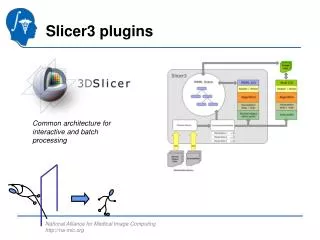
The Slicer3 software • An end-user application for image analysis • An open-source environment for software development • A software platform that is both easy to use for clinical researchers and easy to extend for programmers
Software version Disclaimer It is the responsibility of the user of 3DSlicer to comply with both the terms of the license and with the applicable laws, regulations and rules. Slicer3 is a multi-platform software running on Windows, Linux, and Mac OSX.
Tutorial Dataset The Slicer3minute dataset is composed of an MR scan of the brain and 3D surface reconstructions of anatomical structures. The data are part of the SPL Brain Atlas developed by Talos et al. The atlas is available at: http://www.spl.harvard.edu/publications/item/view/1265
Start Slicer3 Start Slicer: Select Start All Programs Slicer3 3.5.2009-11-06Slicer
GUI Overview Menu Toolbar The Graphical User Interface (GUI) of Slicer3 integrates five components: • the Menu Toolbar • the Module GUI Panel • the 3D Viewer • the Slice Viewer • the Slice and 3D View Controller 3DViewer Module GUI Panel Slice Viewer Slice and 3D View Controller
Slicer Welcome The SlicerWelcome module is the module displayed by default. This module gives an overview of the GUI of Slicer3, and data loading & saving functionalities.
Basics Expand or shrink the GUI panel with the arrows at the frame top, or by clicking and dragging the vertical separator Expand or collapse any sub-panel by clicking on its grey title bar.
Loading a 3D Scene Select File Load Scene from the File menu
Loading a 3D Scene Browse to the location of the Slicer3MinuteDataset directory and select the scene file slicer3minute.mrml Click on Open to load the scene
Loading a 3D Scene Slicer displays a 3D model of the head in the 3DViewer, and anatomical MR slices of the brain in the 2D Slice Viewer. 3D Viewer 2D Slice Viewer
Loading a 3D Scene Left click on the menu Modules and select All Modules to display the list of all 95 modules available for image analysis and 3D visualization.
Loading a 3D Scene To access the Models module, browse through the list of modules or click on the icon in the toolbar button.
Loading a 3D Scene Slicer displays the GUI of the module Models
3D Visualization Position the mouse in the 3D Viewer, hold down the left mouse button and drag to rotate the model.
3D Visualization Click on the Slice Visibility icon to display the Axial Slice in the 3D Viewer
3D Visualization Select the Skin model and turn the opacity of the model from 1.0 to 0.0.
3D Visualization The model of the skull bone and eyeballs appear through the model of the skin in the 3D viewer.
3D Visualization Click on the Slice Visibility icon to display the Coronal Slice in the 3D Viewer
3D Visualization Select the 3D model skull bone in the Model Hierarchy, and select the option Clipping
3D Visualization Browse through the coronal slices to expose the 3D model of the white matter and left and right optic nerves.
3D Visualization Select the hemispheric white matter model in the Model Hierarchy and turn off its visibility.
3D Visualization Slicer displays the optic nerve, optic chiasm and optic tracts overlaid on the MR images of the brain.
3D Visualization Windows/Linux users: Position the mouse in the 3D Viewer, hold down the right mouse button and move the mouse down to zoom in. Mac users: Position the mouse in the 3D Viewer, hold down the apple button and the mouse button and move the mouse down to zoom in.
3D Visualization Slicer3 displays a closer view of 3D anatomical structures overlaid on 2D MR slices
Slicer3 minute tutorial • Slicer3 is an open-source software for image analysis and 3D visualization • Slicer3 core functionalities, available modules and built-in libraries represent more than 2.8 million lines of code • Slicer3 is a multi-institution effort to share the latest advances in image analysis with the scientific and clinical community. http://www.slicer.org/pages/Mailinglist wjp@bwh.harvard.edu
Acknowledgments Harvard Clinical and Translational Science Center National Alliance for Medical Image ComputingNIH U54EB005149 Neuroimage Analysis Center (NAC) National Center for Image-Guided Therapy (NCIGT) Surgical Planning Laboratory, Brigham and Women’s Hospital