Download
1 / 1
10 likes | 75 Vues
A. . B. 100bp Ladder. 2hours. 3hours. 2hours. 3hours. Control. Control. 1hour. 1hour. C. Fig 9. DNA Fragmentation Assay. . Arsenic and Apoptosis in PHLC-1 cells Yeong-Nam Jeong and Dr. Elizabeth Murray Marshall University, Integrated Science and Technology Department. Abstract

E N D
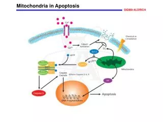
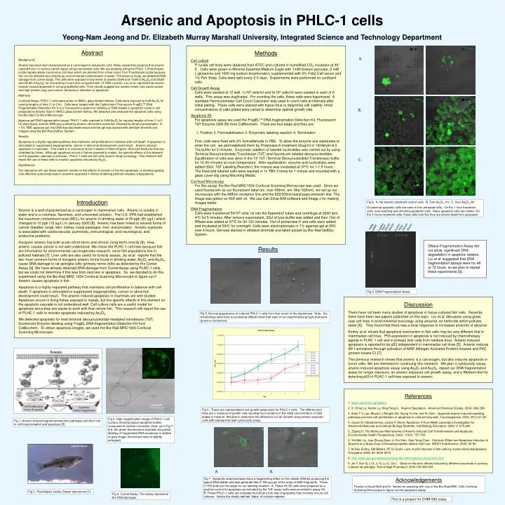
A. B. 100bp Ladder 2hours 3hours 2hours 3hours Control Control 1hour 1hour C. Fig 9. DNA Fragmentation Assay. Arsenic and Apoptosis in PHLC-1 cells Yeong-Nam Jeong and Dr. Elizabeth Murray Marshall University, Integrated Science and Technology Department Abstract Background: Arsenic has been well characterized as a carcinogen in eukaryotic cells. Many researches propose that arsenic caused tumor or various cancer types using mammalian cells. We are studying using the PHLC-1 (Poeciliopsis lucida hepatocellular carcinoma) cell lines which are derived from a liver tumor from Poeciliopsis lucida because fish can be affected very directly by environmental contamination in water. The previous study, we detected DNA damage from comet assay. The cells were exposed in low levels of arsenic (5uM and 10uM of As2O3 and 25uM and 50uM of As2O5) for one and two hours and compared with 1X PBS controls. Lau et al. reported that arsenic trioxide caused apoptosis in rat lung epithelial cells. Their results suggest low arsenic levels may cause cancer and high arsenic may cure cancer, because of induction of apoptosis. Methods: Confocal Assay: PHLC-1 cells were grown in WillCo glass bottom dishes. Cells were exposed to 5uM As2O3 for varying lengths of time (1 to 6 hr). Cells were treated with the Calbiochem Fluorescein-FragELTM DNA Fragmentation Detection Kit. It is a fluorescence system for labeling of DNA breaks in apoptotic nuclei in cell preparations directly fixed in WillCo glass bottom dishes. We detected and analyzed the apoptotic cells using the Bio-Rad Confocal Microscope. Agarose gel DNA fragmentation assay: PHLC-1 cells exposed to 5uM As2O3 for varying lengths of time (1 to 6 hr) were lysed, and the DNA was purified by phenol–chloroform extraction followed by ethanol precipitation. In 1.0% TBE–agarose gel, the DNA was electrophoresed and the gel was stained with ethidium bromide and imaged using the Bio-Rad GelDoc System. Results: Apoptosis is a highly regulated pathway that maintains cell proliferation in balance with cell death. If apoptosis is stimulated or suppressed inappropriately, cancer or abnormal development could result. Arsenic induces apoptosis in mammals. This metal is a commonly found in waters in West Virginia, Ohio and Kentucky that are inhabited by fishes. Although apoptosis occurs in fishes exposed to metals, the specific effects of this element on the apoptotic cascade is unknown. PHLC-1 cells are fish cells used to study toxicology. This research will report the use of these cells to monitor apoptosis induced by As2O3. Significance: Our laboratory will use these research results on the effects of arsenic on the fish apoptosis, to develop speedy, cost-effective cytotoxicity tests to examine apoptosis in fishes inhabiting polluted streams in Appalachia. • Methods • Cell culture • P. lucida cell lines were obtained from ATCC and cultured in humidified CO2 incubator at 30° C. Cells were grown in Minimal Essential Medium Eagle with 1mM Sodium pyruvate, 2 mM L-glutamine and 1500 mg sodium bicarbonate/L supplemented with 5% Fetal Calf serum and 1% Pen Strep. Cells were split every 2-3 days. Experiments were performed on confluent cells. • Cell Growth Assay • Cells were seeded at 12 well. 1x10⁵ cells/ml and 3x10⁴ cells/ml were seeded in each of 6 wells. This assay was duplicated. For counting the cells, these cells were trypsinized. A standard Hemocytometer Cell Count Calculator was used to count cells at intervals after initial plating. These cells were stained with trypan blue to determine cell viability. Initial concentrations of cells plated were varied to determine optimal growth rate. • Apoptosis Kit • For apoptosis assay we used the FragELTM DNA fragmentation Detection Kit, Fluorescent TdT Enzyme (QIA 39) from CalBiochem. There are four steps and they are: • Fixation 2. Permeabilization 3. Enzymatic labeling reaction 4. Termination • First, cells were fixed with 4% formaldehyde in PBS. To allow the enzyme and substrates to enter the cell, we permeabilized them by Proteinase K treatment 20ug/ml in 10mM pH 8.0 Tris buffer for 5 minutes. Enzymatic addition of labeled nucleotides was carried out by using Terminal Deoxynucleotidyl Transferase (TdT) and fluorescein labeled deoxynucleotides. Equilibration of cells was done in the 1X TdT (Terminal Deoxynucleotidyl Transferase) buffer for 10-30 minutes at room temperature. After equilibration, enzyme and nucleotides were • added (60ul TdT Labeling Reaction); the mixture was incubated at 37oC for 1-1.5 hours. The fixed and labeled cells were washed in 1x TBS 3 times for 1 minute and mounted with a glass cover slip using Mounting Media. • Confocal Microscopy • For this assay, the Bio-Rad MRC1024 Confocal Scanning Microscope was used. Since we used fluorescein as our fluorescent label (ex. max 490nm, em. Max 525nm), we set up our microscope with the 488nm excitation line and the 522/329nm band pass emission line. The image was gotten on 40X with oil. We use Carl Zeiss AIM software and Image J for making images better. • DNA Fragmentation • Cells were transferred 5X10⁵ cells/ ml into the Eppendorf tubes and centrifuge at 2000 rpm, 4oC for 5 minutes. After remove supernatant, 20ul of lysis buffer was added and then 10ul of RNase was added at 37oC for 30-120 minutes. 10ul of proteinase K and cells were added and incubated at 50oC for overnight. Cells were electrophoresis in 1% agarose gel at 35V, over 4 hours. Gel was stained in ethidium bromide and taken picture by Bio-Rad GelDoc System. Introduction Arsenic is a well-characterized as a carcinogen in mammalian cells. Arsenic is soluble in water and is a colorless, flavorless, and unscented solution. The U.S. EPA had established the maximum contaminant level (MCL) for arsenic in drinking water of 50 ppb (50 ug/L) which changed to 10 ppb (10 ug/L) in January 2005 [8]. Arsenic has been linked to several forms of cancer (bladder, lungs, skin, kidney, nasal passages, liver, and prostate). Arsenic exposure is associated with cardiovascular, pulmonary, immunological, and neurological, and endocrine problems. Inorganic arsenic has both acute (short-term) and chronic (long-term) toxicity [8]. How arsenic causes cancer is not well understood. We chose the PLHC-1 cell lines because fish are informative for environmental carcinogenesis research, since fish populations live in polluted habitats [7]. Liver cells are also useful for toxicity assays. Jia et al. reports that the two most common forms of inorganic arsenic forms found in drinking water, As2O3 and As2O5, cause DNA damage to rat astroglia cells (primary nerve cells) as detected by the Comet Assay [9]. We have already detected DNA damage from Comet Assay using PLHC-1 cells, but we could not determine if this was from necrosis or apoptosis. So, we decided to do this experiment using the Bio-Rad MRC 1024 Confocal Scanning Microscope to figure out if Arsenic causes apoptosis in fish. Apoptosis is a highly regulated pathway that maintains cell proliferation in balance with cell death. If apoptosis is stimulated or suppressed inappropriately, cancer or abnormal development could result. The arsenic induced apoptosis in mammals are well studied. Apoptosis occurs in living fishes exposed to metals, but the specific effects of this element on the apoptotic cascade is not understood well. Cell culture cells are a useful model for apoptosis since they are easier to work with than whole fish. This research will report the use of PLHC-1 cells to monitor apoptosis induced by As2O3. We detected apoptosis for treat terminal deoxynucleotidyl-mediated transferase (TdT) fluorescent Enzyme labeling using FragEL DNA fragmentation Detection Kit from CalBiochem. To obtain apoptosis images, we used the Bio-Rad MRC1024 Confocal Scanning Microscope. Fig 8. A. No arsenic treatment control cells. B. 5um As2O3 1hr. C. 5um As2O3 3hr Occasional apoptotic cells are seen in the untreated cells. On the 1 hour treatment cells, note blobbing and shrinking apoptotic cells. Many apoptotic cells are visible. On the 3 hours treatment cells, these cells look like they are almost dead from apoptosis. DNase Fragmentation Assay did not show significant DNA degradation in apoptotic ladders. Liu et al. suggested that DNA fragmentation assays were for 48 or 72 hours, so we plan to repeat these experiments [6]. Results Discussion There have not been many studies of apoptosis in tissue cultured fish cells. Recently there have been two papers published on this topic. Liu et al. discusses using grass carp cell lines in environmental toxicology using atrazine, an herbicide which pollutes water [6]. They found that there was a dose response to increased amounts of atrazine. Embry et al. shows that apoptosis mechanism in fish cells may be very different that in mammalian cell lines. P53 expression in apoptosis is not induced by chemotherapy agents in PLHC-1 cell and in primary liver cells from rainbow trout. Arsenic induced apoptosis is reported to be p53 independent in mammalian cell lines [5]. Arsenic induces AP-I activations through activation of MAP (Mitogen Activated Protein) kinases and PKC (protein kinase C) [7]. This previous research shows that arsenic is a carcinogen, but also induces apoptosis in tumor cells. We are interested in continuing this research. We plan a cytotoxicity assay, arsenic induced-apoptosis assay using As2O3 and As2O5, repeat our DNA fragmentation assay for longer exposure, an arsenic exposure cell growth assay, and a Western blot for detecting p53 in PLHC-1 cell lines exposed to arsenic. Fig 5. Normal appearance of cultured PHLC-1 cells from liver tumor of the topminnow. Note, the morphology seen here is somewhat different than that seen in our experimental groups that were grown to confluence. References 1. www.nativefish.org/Gallery 2. X. Chris Le, Xiufen Lu, Xing-Fang Li. Arsenic Spectation. Americal Chemical Society. 2004; 26A-33A 3. Andy T.Y. Lau, Muyai Li, Ronglin Xie, Quing-Yu He, Jen-Fu Chiu. Opposed arsenic-induced signaling pathways promote cell proliferation or apoptosis in cultured lung cells. Carcinogenesis. 2004; 25(1):21-28 4. Susan M. Dibartolomeis, James P. Mone. Apoptosis: A Four-Week Laboratory Investigation for Advanced Molecular and Cellular Biology Students. Cell Biology Education. 2003; 2: 275-295 5. Zigang D. The Molecular Mechanisms of Arsenic-Induced Cell Transformation and Apoptosis. Environmental Health Perspectives. 2002; 110(5): 757-759 6. Xin-Mei Liu, Jian-Zhong Shao, Li-Xin Xian, Xian-Yong Chen. Cytotoxic Effect and Apoptosis Induction of Atrazine in a Grass Carp (Ctenopharyngodon idellus) Cell Line. WILEY InterScience. 2006. 80-89 7. M Rau Embry, SM Billiard, RT Di Giulio. Lack of p53 induction in fish cells by model chemotherapeutics. Oncogene. 2006; 25: 2004-2010 8. http://www.epa.gov/watersecurity/guide/chemicalsensorforarsenic.html 9. Jin Y, Sun G, Li X, Li G, Lu C, Qu L. Study on the toxic effects induced by different arsenicals in primary cultured rat astroglia. Toxicol Appl Pharmacol. 2004;196:396-403. Fig 6. These are representative cell growth assay plots for PHLC-1 cells. The different plot lines are a measure of growth rate resulting from variation in the initial concentration of cells plated in medium. We plan to determine the difference in Cell Growth using arsenic exposed cells with standard 96 well cytotoxicity assay. Fig 2. High magnification image of PHLC-1 cell nucleus showing typical apoptotic bodies composed of nuclear chromatin. Here, as in Fig 7 & 8, the green fluorescence indicates enzymatic labeling of fragmented DNA (scale bar is related to grey image, fluorescent inset is slightly enlarged). Fig 1. Arsenic-induced signal transduction pathways and their role in cell transformation and apoptosis [5]. A. B. Acknowledgements Thanks to David Neff and Dr. Norton for assisting with use of the Bio-Rad MRC 1024 Confocal Scanning Microscope to figure out the apoptosis assay. Fig 7. Apoptotic endonucleases have a fragmenting effect on the cellular DNA by producing the typical DNA ladder and also generate free 3’-OH groups at the ends of DNA fragments. These 3’-OH ends are the target for our labeling reaction. A. These HL-60 cells were prepared as a positive control for apoptosis as indicated by the TdT assay (cells were provided in assay kit). B. These PHLC-1 cells are untreated but show a low rate of apoptosis that normally occurs cell cultures. Notice the clearly defined ‘blebs’ of nuclear material. Fig 3. Poeciliopsis lucida, Desert topminnow [1]. Fig 4. Comet Assay. This assay represents the DNA damages. This is a project for CHM 583 class.