Apoptosis
Apoptosis. Dr Shoaib Raza. Apoptosis . It is a pathway of cell death Induced by tightly regulated suicide program Activated enzyme that degrade cells’ own nuclear DNA, nuclear and cytoplasmic protein Formation of apoptotic bodies that contain Nuclear material and


Apoptosis
E N D
Presentation Transcript
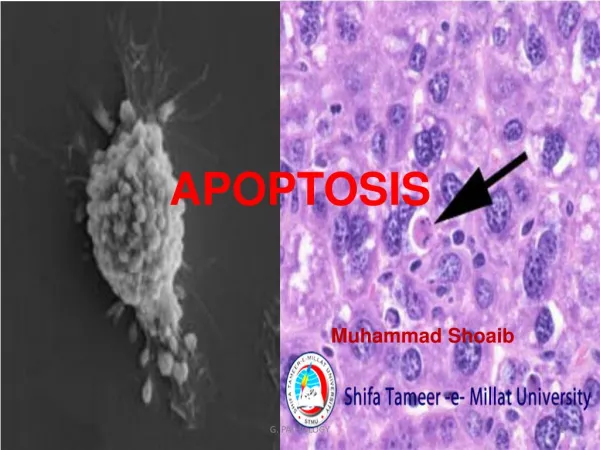
Apoptosis Dr Shoaib Raza
Apoptosis • It is a pathway of cell death • Induced by tightly regulated suicide program • Activated enzyme that degrade cells’ own nuclear DNA, nuclear and cytoplasmic protein • Formation of apoptotic bodies that contain • Nuclear material and • Cytoplasmic organelles and material • No inflammatory response is elicited.
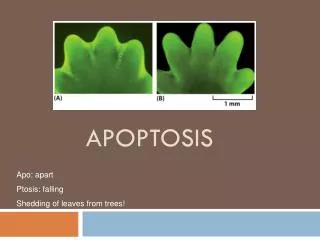
Physiologic examples of Apoptosis • Normal phenomenon that seems to eliminate the cells that are no longer needed • During embryogenesis, metamorphosis, etc • Involution of hormone dependent tissues after hormonal withdrawal • Cell loss in proliferating cell populations • Elimination of potentially harmful self reactive lymphocytes • Death of host cells that have subserved their useful purposes
Pathologic examples of Apoptosis • Eliminate cells that are injured beyond repair without eliciting a host reaction • DNA damage • Accumulation of misfolded protein • Cell death in certain infections (viral) • Viral factors (HIV) • Host factors (HBV) • Pathologic atrophy in parenchymal organs after duct obstruction
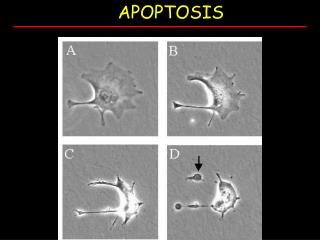
Morphology of Apoptosis • Cell shrinkage • Smaller size, dense cytoplasm, tightly packed organelles • Chromatin condensation • Peripheral aggregation of chromatin, breakup of nucleus • Apoptotic bodies formation • Phagocytosis by macrophages
Biochemical features of Apoptosis • Activation of caspases: • Caspases are proteolytic enzymes present in an inactive form in the cytoplasm • Cysteine proteases that cleaves aspartic acid residue (c asp ases) • Caspases are of two types • Initiator • Initiates and activates other caspases (8,9,10) • Executioner • Executes proteolysis and fragmentation of nucleus (3,6)
Biochemical features of Apoptosis • DNA & protein breakdown • Characteristic breakdown of DNA into large (50,000 – 3000,000) base pairs • Subsequent cleavage of DNA by Ca or Mg dependent endonuclease fragmentation into 180-200 base pairs • Membrane alteration & phagocytosis • Membrane expresses more receptors for phagocytic cells • Phagocytosis is mainly by macrophages • No inflammatory response is elicited
Caspases Activation • Activation of caspases may occur in two ways • Intrinsic (Mitochondrial) Pathway • Pro and anti apoptotic factors are usually present in cell • An imbalance would result in apoptosis • Bcl family of proteins are main regulators • Bcl-2, Bcl-x are main antiapoptotic factors • Bax/Bak channels are major proapoptotic mechanism • Release of Cytochrome-C and other proapoptotic proteins from mitochondria into cytoplasm • Cytochrome-C and Apaf-1 (apoptosis activating factor-1) activates initiators
Extrinsic Pathway • Is activated by • Engagement of plasma membrane death receptor • Fas binds with fasL • Fas associated death domain (FADD) activates • Procaspase-8 is converted into caspase-8 • Initiator sequence begins
Execution Phase • Intrinsic or extrinsic pathway converge at the activation of execution pathway • Important Executioner caspases are caspase-3, caspase-6 • They in turn activate DNAase • Break down of nuclear matrix • Proteolysis of cytoplasmic proteins
Example of Apoptosis • Apoptosis after growth factor deprivation: • Endometrium, lymphocytes, neurons etc. • DNA damage-mediated apoptosis: • Radiation/chemotherapeutic agents induced apoptosis • Apoptosis by TNF family of receptors • Fas-FasL binding • CTL Mediated apoptosis: • Foreign antigen via perforin and granzyme B
Dysregulated Apoptosis • Too little or too much • Defective apoptosis (Increased survival) • Cancer (carcinogenesis) • Autoimmune disorders • Increased apoptosis (Excessive cell death) • Neurogenerative disorders • Infarction and stroke • Death of virus infected cell