Apoptosis
Apoptosis. Apoptosis helps regulate animal cell numbers Is Greek for falling off Most common form of programmed cell death Cellular death is usually proportional to cellular division Billions of cells die in bone marrow and intestine every hour. Introduction. Formation of hands and toes


Apoptosis
E N D
Presentation Transcript
Apoptosis helps regulate animal cell numbers • Is Greek for falling off • Most common form of programmed cell death • Cellular death is usually proportional to cellular division • Billions of cells die in bone marrow and intestine every hour Introduction
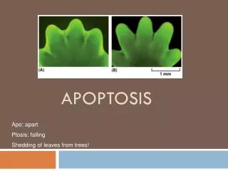
Formation of hands and toes • Others die when structure they provide is no longer needed, like the tail of a tadpole disappearing at metamorphosis. What is the purpose of cell suicide?
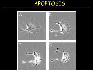
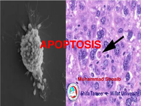
Cells that dies as a result of acute injury typically swell and burst spilling their contents all over neighbor cells • Eruption may trigger a potentially damaging inflammatory response, killing the surrounding cells or damaging them. • In contrast, apoptosis allows the cell to die cleanly without damaging neighbor cells. Apoptosis vs Cell Necrosis
Intrinsic or mitochondrial pathway is initiated from within the cell. • Initiated usually from damage to the DNA or loss of survival factors. • Extrinsic occurs when pro-apoptotic receptors are activated on the cell surface • Receptors cluster and for a death-inducing signal complex(DISC) Intrinsic vs Extrinsic
Machinery involved in apoptosis is similar in all animal cells, involving the caspasefamily of proteases. • Made in inactive form called procaspase • Typically activated by proteolytic cleavage in response to signals that induce apoptosis. • Activated caspases cleave, and thereby activate, other members of procaspasefamily resulting in an amplifying proteolytic cascade. (FIG 18-39 • They also cleave other key proteins like the nuclear lamina which causes the irreversible breakdown of the nuclear lamina. Caspase
Proteolytic cascade is irreversible, destructive and self-amplifying • Therefore, decision to die must be tightly controlled. Important Note
Main proteins that regulate activation of procaspases are members of the Bcl2 family of intracellular proteins. • Some promote activation of apoptosis, leading to cell death while others inhibit apoptosis. Regulation of Intracellular Apoptosis
Activate procaspase indirectly by inducing release of cytochrome c from the mitochondria into cytosol. • Cytochrome c promotes assembly of a seven armed pinwheel structure that recruits specific procaspasemolecules, forming a complex called an apoptosome. • Procaspase molecules become activated within apoptosome triggering caspasecascade that leads to apoptosis (FIG 18-40) BAK and BAX Proteins
Bcl2 itself and other members of the family act to inhibit procaspase activation and apoptosis • One way of doing this is by blocking the ability of Bax and Bak to release cytochrome c from mitochondria. • Protein Bad promotes apoptosis by binding to Bcl2 and the other death-suppressing proteins of the family to block its activity. Bcl2
Cancer cells are less likely to kill themselves. • This is occurs because of a mutation in the P53 gene. • P53 gene normally acts as checkpoint to have cell cease dividing or to die by apoptosis when their DNA is damage Cancer
Research Apoptosis. (2013). Retrieved December 1, 2013, from Gentech: http://www.biooncology.com/research- education/apoptosis/pathways • Alberts, B. (2002). Molecular Biology of the Cell. • Alberts, B. (2010). Essential Cell Biology. New York: Garland Science, Taylor & Francis Group. • Azad, A. (2013). BAK multimerization for apoptosis, but not bid binding, is inhibited by negatively charged residue in the BAK hydrophobic groove. Molecular Cancer. References