Understanding Apoptosis: Cell Death Mechanism and Importance
Learn about apoptosis, a controlled cell death process crucial for development and homeostasis, but improper regulation contributes to diseases like cancer. Discover the characteristics, morphology, and differences from necrosis. Explore the mechanisms, stimuli, and factors involved in apoptotic cell death.


Understanding Apoptosis: Cell Death Mechanism and Importance
E N D
Presentation Transcript
In the human body about 100,000 cells are produced every second by mitosis and a similar number die by apoptosis !!! APOPTOSIS
Definition Apoptosis is a peculiar well controlled individual cell death that is caspase mediated and leads to fragmentation of the cell and organelles into numerous small buds, which are then engulfed by macrophages without surrounding inflammation.
Importance of Apoptosis 1) Crucial for embryonic development -Errors in Apoptosis can lead to Birth Defects 2) Important for maintaining homeostasis - Cell death is balanced with mitosis to regulate cell number. 3) Improper regulation contributes to human disease - Neurodegenerative diseases Parkinson’s Alzheimer’s -Cancer - Autoimmune diseases e.g. (diabetes type I) - Viral diseases
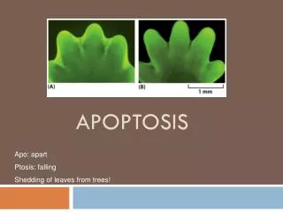
Characteristics • It is a process that occurs in almost all living creatures since their early stages of embryological development. • It is an active cytological process in which energy is consumed (ATP dependent) • It is programmed or controlled by genetic protocol or program (control of enzymes, cell membrane surface proteins & cytoplasmic molecules, signal transduction, gene expression) • It may be triggered by intrinsic or extrinsic stimuli
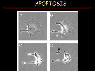
Morphology • Cell shrinkage (condensation of cytoplasm) due to breakdown of cytoskeleton • Breakdown of mitochondria; release of cytochrome C • Nuclear condensation • Nuclear fragmentation • Cell membrane blebbing • Fragmentation; apoptotic body formation: membrane-bound cellular fragments, which often lack nuclei • Phagocytosis
How Apoptosis Differs from Necrosis? • Apoptosis is intrinsically controlled, necrosis is not • Apoptosis is more rapid (12-24 hours) than necrosis • Apoptosis is induced by endogenous or exogenous stimuli, necrosis is always induced by exogenous harms • Apoptosis is limited to single or few cells at a time, and occurs among healthy cell population, necrosis is usually more extensive & occurs in tissue exposed to injuries • Cell cytoplasm shrinks in apoptosis and swells in necrosis. • Nucleosomes of apoptotic cells are 180 bp fragments, contrary to the irregular ones in necrosis • Apoptosis has no inflammation, while necrosis leads to liberation of pro-inflammatory mediators • Apoptosis has no systemic manifestations contrary to most inflammations
Mechanism I. Four stages of apoptosis have been defined: i. Committment to death by extracellular or intracellular triggers/signals ii. Cell killing (execution) by activation of intracellular proteases (caspases) iii. Engulfment of cell corpse by other cells iv. Degradation of the cell corpse within the lysosomes of phagocytic cells
II. Stimuli for Apoptotic Cell Death in Mammals i. Growth factor deficiencies ii. Ionizing radiation/ viral infection iii. Free radical toxicity iv. Death receptor activation (such as Fas or CD95 triggering) v. Metabolic or cell cycle perturbation
Death Factors Definition: cytokines that activate an apoptosis program by binding to their specific receptor. Typical examples of death factors are: • Fasligand, • TNF (tumor necrosis factor) and • TRAIL (TNF-related apoptosis-inducing ligand). - Apoptosis can also be induced by cytotoxic T-lymphocytes using the enzyme granzyme.
III. Activation of Caspase cascade i. Various stimuli described above eventually activate the executioner (caspase) cascade ii. At least 14 different caspases exist in human cells iii. Caspase cascades are apparently required for complete execution
The extrinsic (death receptor-initiated) pathway of apoptosis, illustrated by the events following Fas engagement. FADD= Fas associated death domain.
Cell injury e.g., radiation , toxins, free radicals Apoptotic body Cytoplasmic bleb
The intrinsic (mitochondrial) pathway of apoptosis. Death agonists cause changes in the inner mitochondrial membrane, resulting in the mitochondrial permeability transition (MPT) and release of cytochrome c and other pro-apoptotic proteins into the cytosol, which activate caspases. AIF= Apoptosis inhibitory factor; IAPs= Inhibitors of apoptosis proteins; Apaf-1= apoptosis protease activating factor
Stimuli that activate intrinsic pathway • growth factor withdrawal, DNA damage, excessive growth signaling (oncogenes), viral infection and cell cycle abnormalities)
Caspases are central initiators and executioners of apoptosis • The term caspases is derived from cysteine-dependent aspartate-specific proteases: their catalytical activity depends on a critical cysteine-residue within a highly conserved active-site pentapeptide QACRG, and the caspases specifically cleave their substrates after Asp residues. • Initiator caspases include: 2, 8, 9, 10 • Execution caspases include: 3, 6, 7
The caspase cascade can be activated by: • Granzyme B released by cytotoxic T lymphocytes which is known to activate caspase-3 and -7; • death receptors (like FAS, TRAIL receptors and TNF receptor) which can activate caspase-8 and -10; and • the apoptosome, regulated by cytochrome c and the Bcl-2 family, which activates caspase-9.
Extrinsic apoptosis pathway (e.g. procaspase-8 is recruited by its DEDs to the death inducing signalling complex (DISC), a membrane receptor complex formed following to the ligation of a member of the tumor necrosis factor receptor (TNFR) family [Sartorius, 2001]. When bound to the DISC, several procaspase-8 molecules are in close proximity to each other and therefore are assumed to activate each other by autoproteolysis [Denault, 2002].
Intrinsic apoptosis pathway Intrinsic apoptosis pathway involve procaspase-9 which is activated downstream of mitochondrial proapoptotic events at the so called apoptosome, a cytosolic death signalling protein complex that is formed upon release of cytochrome c from the mitochondria [Salvesen, 2002]. In this case it is the dimerization of procaspase-9 molecules at the Apaf-1 scaffold that is responsible for caspase-9 activation [Denault, 2002]. Once the initiator caspases have been activated, they can proteolytically activate the effector procaspases-3, -6, and -7 which subsequently cleave a specific set of protein substrates, including procaspases themselves, resulting in the mediation and amplification of the death signal and eventually in the execution of cell death with all the morphological and biochemical features usually observed [Earnshaw, 1999].
Bcl-2 • Bcl2 was the first apoptosis-related gene that was recognized to play a role in tumorigenesis, and indeed, Bcl-2 is overexpressed in a variety of cancers, contributing to cancer cell survival through direct inhibition of apoptosis.
BCL-2 • BCL-2 is a human proto-oncogene located on chromosome 18. • Its product is an integral membrane protein (called Bcl-2) located in the membranes of the endoplasmic reticulum (ER), nuclear envelope, and in the outer membrane of the mitochondria. • The gene was discovered as the translocated locus in a B-cell leukemia (hence the name). This translocation is also found in some B-cell lymphomas.