Elbow and Hand
PTA 106 Fall 2008. Elbow and Hand . Chanel Kyle Sandra. Surface Anatomy. Lateral Epicondyle Medial Epicondlye Olcranon Ulnar Styloid Process Cubital Fossa Site of Median Nerve Tendon of Palmaris Longus Tendon of Flexor Carpi Radialis Distal Wrist Crease. Anatomical Snuff Box

Elbow and Hand
E N D
Presentation Transcript
PTA 106 Fall 2008 Elbow and Hand Chanel Kyle Sandra
Surface Anatomy • Lateral Epicondyle • Medial Epicondlye • Olcranon • UlnarStyloid Process • CubitalFossa • Site of Median Nerve • Tendon of Palmaris Longus • Tendon of Flexor Carpi Radialis • Distal Wrist Crease
Anatomical Snuff Box • Thenar Eminance • Hypothenar Eminence
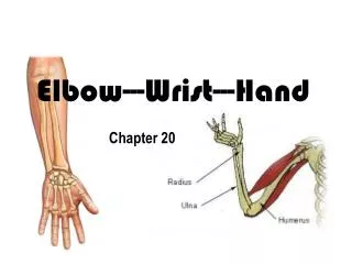
The Humerus • Medial Epicondyle • Lateral Epicondyle • Capitulum- on lateral edge of condyle • Trochlea- medial and mid section of condyle • Coronoid Fossa • Olecranon Fossa
Ulna • Olecranon Process • Trochlear Notch • Coronoid Process • Radial Notch • Ulnar Tuberosity • Styloid Process
Radius • Head • Neck • Radial Tuberosity • Ulnar Notch • Styloid Process
Wrist/Hand • Scaphoid, Lunate, Triquetrum, Pisiform, Trapezium, Hamate, Trapezoid, Capitate • Metacarpals: Numbered 1-5, starting at thumb • Phalangeal: Numbered 1-5 • Distal, Middle, Proximal
LIGAMENTS OF THE ELBOW (HUMEROULNAR JT) • A. Ligaments • Elbow • Articular Capsule • Radial anular ligament • Interosseous membrane • Ulnar collateral ligament • Radial collateral ligament • Hand • Palmar aponeurosis • Common Flexor sheath • Flexor retinaculum • B. Bursae • Subcutaneous olecranon bursa • Subtendinous olecranon bursa • C. Cartilage, articular cartilage • D. Articular Capsule • Synovial membrane • Fibrous layer
LIGAMENTS OF THE ELBOW (HUMEROULNAR AND HUMERORADIAL JOINTS) • Articular Capsule • Radial Anular ligament • Interosseous membrane • Ulnar collateral ligament • Radial collateral ligament • All provide strength and support to the joint as do the surrounding muscles • Two ligaments found in the elbow joint are: • the ulnar collateral ligament and the radial collateral ligament. • They are strong, fan shaped condensations of the fibrous joint capsule
LIGAMENTS OF THE ELBOW (HUMEROULNAR JT) • Movements of the elbow joint • Flexion and extension occur at the elbow joint • The long axis of the fully extended ulna makes an angle of approximately 170° with the long axis of the humerus • Called the carrying angle for the way the forearm angles away from the body when something is carried • The obliquity of the angle is more pronounced in women than in men
LIGAMENTS OF THE ELBOW (HUMEROULNAR AND PROXIMAL RADIOULNAR JT) • Articular capsule • A sac enclosing a joint, formed by an outer fibrous membrane and an inner synovial membrane. Also called joint capsule • The synovial fluid nourishes the fibrocartilage and lubricates the joint surface
LIGAMENTS OF THE ELBOW (HUMEROULNAR JT) • Weak anteriorly and posteriorly, the capsule strengthened on each side by the ulnar and radial collateral ligaments • The fibrous layer of the capsule is continuous with the fibrous layer of the elbow joint • It attaches to the humerus at the margins of the lateral and medial ends of the articular surfaces of the capitulum and trochlea • Fig 6.35
LIGAMENTS OF THE ELBOW (HUMEROULNAR JT) • Radial anular ligament • Encircles and holds the head of the radius in the radial notch of the ulna • Forms the proximal radioulnar joint • Allows pronation and supination of forearm • In pronation/supination it is the radius that rotates • Figure 6.33E, 6.36C
LIGAMENTS OF THE ELBOW (HUMEROULNAR JT) • Ulnar collateral ligament • Located on the medial side of the joint, it extends from the medial epicondyle of the humerus to the proximal portion of the ulna • Prevents excessive abduction of the elbow joint
LIGAMENTS OF THE ELBOW (HUMEROULNAR JT) • Radial collateral ligament • Located on the lateral side of the joint, extending from the lateral epicondyle of the humerus to the head of the radius. • Prevents excessive adduction of the elbow joint.
LIGAMENTS OF THE ELBOW (HUMEROULNAR JT) • Interosseous membrane • Connects the shafts of the ulna and the radius throughout most of their length • Classified as a fibrous joint, or syndesmosis • Note: The ulnar and radial collateral ligaments of the proximal humeroulnar joint are not to be confused with the ligaments of the same name at the distal radioulnar joint • Fig 6.20A,B
LIGAMENTS OF THE HAND • Fascia of the palm is continuous with the antebrachial fascia, and the fascia of the dorsum of the hand • Palmar aponeurosis • Common Flexor Sheath • Flexor retinaculum
LIGAMENTS OF THE HAND • Palmar aponeurosis • Strong, well-defined, central part of the palmar fascia • Covers soft tissues and overlies long flexor tendons • Proximal end is continuous with the flexor retinaculum and palmaris longus tendon • Fig 6.25C
LIGAMENTS OF THE HAND • Palmar aponeurosis • The end distal to apex forms four longitudinal bands that radiate from the apex • These attach distally to the bases of the proximal phalanges • Forming the fibrous digital sheaths of individual digits
LIGAMENTS OF THE HAND • Flexor Retinaculum • Or transverse carpal ligament • Continuous with the antebrachial fascia • Fibrous band that extends between the anterior prominences of the outer carpal bones
LIGAMENTS OF THE HAND • Flexor retinaculum • and converts the anterior concavity of the carpus into the carpal tunnel • Through which the flexor tendons and median nerve pass
LIGAMENTS OF THE HAND • Common Flexor Sheath • Deep to the Flexor retinaculum • Together with the digital sheaths enables the tendons to slide freely past each other during movement
BURSAE OF THE HUMEROULNAR JT AND HAND • Bursa • closed sacs containing fluid which prevent friction, and enable structures to move freely over one another • Subcutaneous olecranon bursa-Located in the subcutaneous connective tissue over the olecranon • Subtendinous olecranon bursa-Located between the olecranon and triceps tendons, just proximal to its attachment
CARTILAGE • Articular cartilage • Caps the articulating surfaces of bones participating in a synovial joint • Provides a smooth, low-friction gliding surface • Avascular, nourished by diffusion (synovial fluid)
Biceps Brachii O: Scapula: Long Head: Supraglenoid Tubercle; Short Head: Coracoid Process. I: Radial Tuberosity of Raidus. A: Elbow Flexion, Forearm Supination. I: Musculocutaneous Nerve Vascular: Brachial Artery
Triceps Brachii O: Long Head- Infraglenoid Tubercle of Scapula,Lateral Head- Inferior to Great Tubercle on Posterior HumerusMedial Head- Posterior Surface of Humerus I: Olecranon Process of Ulna A: Elbow Extension I: Radial Nerve Vascular: Deep Brachial Artery
Coracobrachialis O: Apex of the Coracoid Process I: Middle of the medial Surface and border of the Humerus. A: Flexes and adducts the arm. I: Musculocutaneous Nerve (C6 and C7) Vascular: Brachial Artery
Brachialis O: Distal Half of Humerus, Anterior Surface I: Coronoid Process and Ulnar Tuberosity of the Ulna A: Elbow Flexion I: Musculocutaneous Nerve Vascular: Brachial Artery
Brachioradialis O: Lateral Supracondylar Ridge on the Humerus I: Styloid Process of the Radius A: Elbow Flexion I: Radial Nerve Vascular: Radial Artery
Supinator O: Later Epicondyle of Humerus and adjacent Ulna I: Anterior Surface of the Proximal Radius A: Forearm Supination I: Radial Nerve Vascular: Recurrent Interosseous Artery
1) Pronator Teres O: Medial Epicondyle of Humerus and Coranoid Process of Ulna I: Lateral aspect of Radius at its midpoint A: Forearm Pronation, Assistive in elbow flexion I: Median Nerve Vascular: Ulnar Artery 2) Pronator Quadratus O: Distal Fourth of Ulna I: Distal Forth of Radius A: Forearm Pronation I: Median Nerve Vascular: Anterior Interosseous Artery
Flexor Carpi Radialis O: Medial Epicondyle of the Humerus I: Base of Second and Third Metacarpals A: Wrist Flexion, Radial Deviation I: Median Nerve Vascular: Radial and Ulnar Arteries
Flexor Carpi Ulnaris O: Medial Epicondyle of Humerus I: Pisiform and base of Fifth Metacarpal A: Wrist Flexion, Ulnar Deviation I: Ulnar Nerve Vascular: Ulnar Artery
Extensor Carpi Radialis Longus O: Supracondylar Ridge of Humerus I: Base of Second Metacarpal A: Wrist extension, Radial Deviation I: Radial Nerve Vascular: Radial Artery
Extensor Carpi Radialis Brevis O: Lateral Epicondyle of Humerus I: Base of Third Metacarpal A: Wrist Extension I: Radial Nerve Vascular: Radial Artery
Extensor Digitorum O: Lateral Epicondyle of Humerus I: Base of distal Phalanx of the Second-Fifth Fingers A: Extends all three joints of the Fingers I: Radial Nerve Vascular: Recurrent Interosseous Artery
Extensor Carpi Ulnaris O: Lateral Epicondyle of Humerus I: Medial Side of Base of 5th Metacarpal A: Extends and Adducts Wrist I: Deep Radial Nerve Vascular: Ulnar Artery
Flexor Digitorum Superficialis O: Common Flexor tendon, Coronoid Process and Radius I: Sides of the Middle Phalanx of the Four Fingers A: Flexes MP and PIP joints of the Fingers I: Median Nerve Vascular: Ulnar Artery
Flexor Digitorum Profundus O: Upper three-fourths of Ulna I: Distal Phalanx of the Four Fingers digits (2-5) A: Flexes all three joints of the Fingers I: Median and Ulnar Nerves Vascular: Ulnar Artery
Flexor Pollicis Longus O: Radius, Anterior Surface I: Distal Phalanx of Pollex A: Flexes all joints of the Pollex or Thumb I: Median Nerve Vascular: Radial Artery
Abductor Pollicis Longus O: Posterior radius, Interosseous Membrane, Middle Ulna I: Base of the First Metacarpal A: Abducts Pollex I: Radial Nerve Vascular: Posterior Interosseous Artery
Extensor Digiti Minimi O: Lateral Epicondyle of Humerus I: Base of Distal Phalanx of Fifth Finger A: Extends all joints of Fifth Finger I: Radial Nerve Vascular: Recurrent Interosseous Artery
Extensor Pollicis Brevis O: Posterior Distal Radius I: Base of the Proximal Phalanx of Pollex A: Extends MP joint of Thumb I: Radial Nerve Vascular: Posterior Interosseous Artery
Extensor Pollicis Longus O: Middle Posterior Ulna and Interosseous Membrane I: Base of Distal Phalanx of Pollex A: Extends MP and IP joints of the Thumb I: Radial Nerve Vascular: Posterior Intercosseous Artery
Palmaris Longus O: Medial Epicondyle of Humerus I: Palmar Fascia A: Assistinve in Wrist Flexion I: Median Nerve Vascular: Ulnar Artery
Carpal Tunnel • How Is It Caused? • Predisposition • Trauma, Injury, Swelling, Spain, Fracture. • Hormones • Overactivity of pituitary, Hypothyroidism • Mechanical Problems • Work stress, vibrating hand tools • Development of Cyst or Tumor in Canal • Some times there is no origin • What Is It? • Happens when the median nerve becomes squeezed or pressed at the wrist • Sometimes caused by thickening of irritated tendons that go through the tunnel. • The result being pain that goes it to the hand and can even radiate up in to the forearm. • Most common of the entrapment neuropathies where the peripheral nerves are traumatized or compressed.
Symptoms • Frequent burning, tingling, itching, numbness in palm of hand and fingers. • Fingers feel worthless and swollen. • Decreased grip strength; difficult to form a fist, grasp small objects, • Can differentiate between hot and cold. • Treatment • Surgical • Open Release • Endoscopic • Non-Surgical • Drugs:NSAID, Corticosteroids, and Vitamin B6 • Exercise: Stretching and strenthening • Alternative: acupuncture, yoga and chiropractic services
Tennis Elbow “Lateral Epicondylitis” • Causes • Tennis or any racket sport • Anything that involves extending your wrist or rotating his forearm, such as twisting a screwdriver or lifting a heavy object with your palm down. • With age irritation becomes inflamed more easily. • What is Tennis Elbow? • Is inflammation around the lateral epicondyle • It occurs when the muscle attachment, tendons, become irritated.
Symptoms • Pain that radiates from epicondyle in to forearm and wrist. • Pain with extension of wrist. • Forearm weakness. • Painful grip with activities such as shaking hands and turning door knobs. • Inability to hold objects such as coffee cups. • Treatment • Use of a brace to let muscles rest. • Corticosteroid injections. • PT: Stretching and ROM exercises. • Surgery is Rare