Download
1 / 21
250 likes | 617 Vues
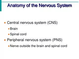
Chapter Four Anatomy of the Nervous System. Divisions of the Vertebrate Nervous System. Central Nervous System-the brain and the spinal cord Peripheral Nervous System-the nerves outside the brain and spinal cord Two Division of the PNS

E N D
Divisions of the Vertebrate Nervous System Central Nervous System-the brain and the spinal cord Peripheral Nervous System-the nerves outside the brain and spinal cord Two Division of the PNS Somatic Nervous System-the nerves that convey messages from the sense organs to the CNS and from the CNS to the muscles and glands Autonomic Nervous System-a set of neurons that control the heart, the intestines, and other organs
Figure 4.1 The human nervous systemBoth the central nervous system and the peripheral nervous system havemajor subdivisions. The closeup of the brain shows the right hemisphereas seen from the midline.
The Nervous System The Spinal Cord-part of the CNS found within the spinal column The spinal cord communicates with the sense organs and muscles below the level of the head Bell-Magendie Law-the entering dorsal roots carry sensory information and the exiting ventral roots carry motor information to the muscles and glands Dorsal Root Ganglia-clusters of neurons outside the spinal cord
Figure 4.3 Diagram of a cross section through the spinal cordThe dorsal root on each side conveys sensory information to the spinal cord; the ventral root conveys motor commands to the muscles.
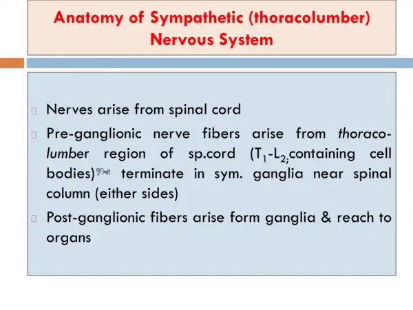
Sympathetic-prepares the body for arousal Ex: increased breathing, increased heart rate, decreased digestive activity Form chain of ganglia just outside spinal cord Short preganglionic axons release norepinephrine Long postganglionic axons release norepinephrine Parasympathetic-facilitates vegetative, nonemergency responses by the body’s organs Ex: increase digestive activity, activities opposing sympathetic system Consists of cranial nerves and nerves from sacral spinal cord Long preganglionic axons extend from the spinal cord to parasympathetic ganglia close to each internal organ; release norepinephrine Shorter postganglionic fibers then extend from the parasympathetic ganglia in the organs; release acetylcholine Autonomic Nervous System
The Brain The Hindbrain/rhombencephalon Posterior part of brain Medulla-controls vital reflexes like breathing, heart beat, etc Pons-Area where many axons cross from one side of the brain to the other Reticular formation-control motor areas of the spinal cord and sends output to cerebral cortex increasing arousal and attention Raphe system-sends axons to much of the forebrain, increasing or decreasing the brain’s readiness to respond to stimuli Cerebellum-control movement, shifts of attention, balance and coordination
The Brain The Midbrain-middle of the brain Tegmentum-”roof or covering” Nuclei for third and fourth cranial nerves Parts of Reticular formation Extensions of the pathways between the forebrain and the spinal cord or hindbrain Tectum-”roof” Superior Colliculus & Inferior Colliculus-important in routes of sensory information
Figure 4.8 The human brain stemThis composite structure extends from the top of the spinal cord into the center of the forebrain. The pons, pineal gland, and colliculi are ordinarily surrounded by the cerebral cortex.
The Brain The Forebrain-most anterior and most prominent part of the mammalian brain Thalamus Part of the Diencephalon Center of forebrain Relay Station for Sensory Information Hypothalamus Part of Diencephalon Regulates homeostasis, sexual behavior, fighting, feeding Pituitary Gland Endocrine gland attached to the base of the hypothalamus
Figure 4.10 The limbic system is a set of subcortical structures that form a border (or limbus) around the brain stem
The Brain Forebrain Cont’d Basal Ganglia Responsible for motor behavior, some memory and emotional expression Basal Forebrain Located on the dorsal surface of the forebrain Received input from the hypothalamus and basal ganglia Send axons to cerebral cortex Important in arousal, wakefulness, and attention Hippocampus Located between thalamus and cerebral cortex Critical for the formation of new memory
Figure 4.14 The basal gangliaThe thalamus is in the center, the basal ganglia are lateral to it, and the cerebral cortex is on the outside.
The Brain The Ventricles-Assists in cushioning the brain Central Canal-fluid-filled channel in the center of the spinal cord Ventricles-four fluid-filled cavities within the brain CSF-clear fluid similar to blood plasma Formed in choroid plexus Flows from lateral to third to fourth ventricle to central canal or between meninges Meninges-membranes that surround the brain and spinal cord
Figure 4.16 The cerebral ventriclesDiagram showing positions of the four ventricles.
The Cerebral Cortex Organization of the Cerebral Cortex Contains six distinct layers of cells Organized into columns-cells with similar properties; arranged perpendicular to the laminae Cells within a given column have similar or related properties
The Lobes The Occipital Lobe-posterior end of cortex Contains primary visual cortex The Parietal Lobe-between occipital love the central sulcus Contains the primary somatosensory cortex-receiving touch sensation, muscle-stretch information and joint position information The Temporal Lobe-lateral portion of each hemisphere, near the temples Contains targets for audition, essential for understanding spoken language, complex visual processes, emotional and motivational behaviors The Frontal Lobe-extends from the central sulcus to the anterior limit of the brain Contains Primary Motor Cortex-fine movements Contributes to shifting attention, planning of action, delayed response tasks as examples
Figure 4.20 Some major subdivisions of the human cerebral cortexThe four lobes: occipital, parietal, temporal, and frontal.
Brain Function How Do the Pieces Work Together? Does the Brain Operate as a Whole or a Collection of Parts? Each brain area has a function but it can’t do much by itself The Binding Problem The question of how the visual, auditory, and other areas of your brain influence on another to produce a combined perception of the single object Synchronized neural activity?