Download
1 / 58
580 likes | 882 Vues
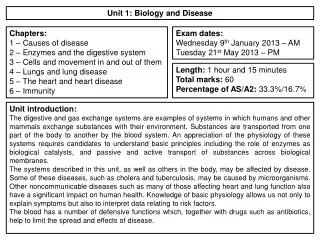
Unit 1: Biology and Disease. Chapters: 1 – Causes of disease 2 – Enzymes and the digestive system 3 – Cells and movement in and out of them 4 – Lungs and lung disease 5 – The heart and heart disease 6 – Immunity. Exam dates: Wednesday 9 th January 2013 – AM Tuesday 21 st May 2013 – PM .

E N D
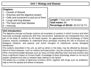
Unit 1: Biology and Disease Chapters: 1 – Causes of disease 2 – Enzymes and the digestive system 3 – Cells and movement in and out of them 4 – Lungs and lung disease 5 – The heart and heart disease 6 – Immunity Exam dates: Wednesday 9th January 2013 – AM Tuesday 21st May 2013 – PM Length: 1 hour and 15 minutes Total marks: 60 Percentage of AS/A2: 33.3%/16.7% Unit introduction: The digestive and gas exchange systems are examples of systems in which humans and other mammals exchange substances with their environment. Substances are transported from one part of the body to another by the blood system. An appreciation of the physiology of these systems requires candidates to understand basic principles including the role of enzymes as biological catalysts, and passive and active transport of substances across biological membranes. The systems described in this unit, as well as others in the body, may be affected by disease. Some of these diseases, such as cholera and tuberculosis, may be caused by microorganisms. Other noncommunicable diseases such as many of those affecting heart and lung function also have a significant impact on human health. Knowledge of basic physiology allows us not only to explain symptoms but also to interpret data relating to risk factors. The blood has a number of defensive functions which, together with drugs such as antibiotics, help to limit the spread and effects of disease.
Unit 1: Chapter 1: Causes of Disease 1.1 Pathogens: What are pathogens? How do pathogens enter the body? How do pathogens cause disease? Key words: damage; infection; microorganisms; pathogens; toxins; What is a pathogen? How do pathogens enter the body? How do pathogens cause disease?
Unit 1: Chapter 1: Causes of Disease 1.2 Data and disease: How are data on disease interpreted and analysed? What is correlation and what does it mean? How is causal link established? Key words: causal link; correlation; How are data on disease interpreted and analysed? What is correlation and what does it mean? How is causal link established?
Unit 1: Chapter 1: Causes of Disease 1.3 Lifestyle and health: What is risk? How is risk measured? What factors affect the risk of contracting cancer? Key words: blood cholesterol; cancer; carcinogenic; diet; emphysema; high blood pressure; obesity; osteoarthritis; physical activity; smoking; sunlight; What is risk? How is risk measured? What factors affect the risk of contracting cancer?
Unit 1: Chapter 1: Causes of Disease Exam questions Other than bacteria name one pathogen: (1 mark) Several diseases are caused by inhaling asbestos fibres. Most of these diseases result from the build up of these tiny asbestos fibres in the lungs. One of these diseases is asbestosis. The asbestos fibres are very small and enter the bronchioles and alveoli. They cause the destruction of phagocytes and the surrounding lung tissue becomes scarred and fibrous. The fibrous tissue reduces the elasticity of the lungs and causes the alveolar walls to thicken. One of the main symptoms of asbestosis is shortness of breath caused by reduced gas exchange. People with asbestosis are at a greater risk of developing lung cancer. The time between exposure to asbestos and the occurrence of lung cancer is 20–30 years. Give two ways in which a pathogen may cause disease: 1) 2) (2 marks) Scientists who investigate disease may look at risk factors. What is a risk factor? (1 mark) Doctors did not make the link between exposure to asbestos and an increased risk of developing lung cancer for many years. Use information in the passage to explain why. (1 mark)
Unit 1: Chapter 1: Causes of Disease Exam questions positive correlation between the number of cases of asthma and the concentration in the air of substances from vehicle exhausts (1 mark) a negative correlation between the number of cases of asthma and the concentration in the air of substances from vehicle exhausts (1 mark) The scientists concluded that substances in the air from vehicle exhausts did not cause the increase in asthma between 1976 and 1980. Explain why. (3marks)
Unit 1: Chapter 2: Enzymes and the digestive system Key words: absorption; assimilation; carbohydrase;egestion; hydrolase; hydrolysis; large intestine; lipase;oesophagus; pancreas;protease; rectum; salivary glands; small intestine; stomach; 2.1 Enzymes and digestion: What are the structure and function of the major parts of the digestive system? How does the digestive system break down food both physically and chemically? What is the role of enzymes in digestion? Label the parts of the digestive system and explain the function of each part: State what chemical and physical digestion are and where take place
Unit 1: Chapter 2: Enzymes and the digestive system 2.2 Carbohydrates – monosaccharaides: How are large molecules like carbohydrates constructed? What is the structure of a monosaccharide? How would you carry out the Benedict’s test for reducing and non-reducing sugars? Key words: Benedict’s test; carbohydrate; monomer; monosaccharide; Draw the monomer α-glucose: Explain how to carry out the Benedict's test: Label the tubes below to show the result: How are large molecules like carbohydrates constructed?
Unit 1: Chapter 2: Enzymes and the digestive system 2.3 Carbohydrates – disaccharides and polysaccharides: How are monosaccharaides linked together to form disaccharides? How are α-glucose molecules linked to form starch? What is the test for non-reducing sugars? What is the test for starch? Key words: cellulose; condensation; disaccharide; glycogen; glycosidic bond; iodine/KI test; polymers; polysaccharide; starch; Glucose links to glucose to form: Glucose links to fructose to form: Glucose links to galactose to form: Draw the formation of maltose, name the bond formed and the type of reaction: Draw the breaking of sucrose and name the type of reaction: What is the test for non-reducing sugars, and what results would you expect? What is the test for starch, and what results would you expect?
Unit 1: Chapter 2: Enzymes and the digestive system 2.4 Carbohydrate digestion: How does salivary amylase act in the mouth to hydrolyse starch? How is starch digestion completed in the small intestine? How are the disaccharides digested? What is lactose intolerance? Key words: amylase; maltase; lactase;pancreatic amylase; salivary amylase; sucrase How is sucrose digested? How is lactose digested? Label the parts of the digestive system; the enzymes they produce and explain their role in the digestion of starch: What is lactose intolerance?
Unit 1: Chapter 2: Enzymes and the digestive system Key words: alpha-helix; amino acid; β-pleated sheet;biuret test;dipeptide; disulphide bonds; ionic bonds; hydrogen bonds; peptide bond; polymerisation; polypeptide; primary structure; protein; quaternary structure; secondary structure; tertiary structure; 2.5 Proteins: How are amino acids linked to for polypeptides – the primary structure of proteins? How are polypeptides arranged to form the secondary structure and then the tertiary structure of a protein? How is the quaternary structure of a protein formed? How are proteins identified? Draw and label an amino acid: Label the diagram to show the formation of a polypeptide bond: What is the test for proteins and what results would you expect?
Unit 1: Chapter 2: Enzymes and the digestive system Key words: alpha-helix; amino acid; β-pleated sheet;biuret test;dipeptide; disulphide bonds; ionic bonds; hydrogen bonds; peptide bond; polymerisation; polypeptide; primary structure; protein; quaternary structure; secondary structure; tertiary structure; 2.5 Proteins: How are amino acids linked to for polypeptides – the primary structure of proteins? How are polypeptides arranged to form the secondary structure and then the tertiary structure of a protein? How is the quaternary structure of a protein formed? How are proteins identified? Draw the quaternary structure of a protein: Draw the primary structure of a protein: Draw the tertiary structure of a protein: Draw the secondary structure of a protein:
Unit 1: Chapter 2: Enzymes and the digestive system 2.6 Enzyme action: How do enzymes speed up chemical reactions? How does the structure of enzyme molecules relate to their function? What is the lock and key model of enzyme action? What is the induced-fit model of enzyme action? Key words: activation energy; catalyst; enzyme; enzyme-substrate complex; induced fit; lock and key; substrate; Draw a diagram to explain the lock and key model of enzyme action: How does an enzyme’s structure relate to it’s function? Draw a diagram to explain the induced-fit model of enzyme action: Draw a sketch graph to show how enzymes speed up a reaction:
Unit 1: Chapter 2: Enzymes and the digestive system 2.7 Factors affecting enzyme action: How is the rate of an enzyme-controlled reaction measured? How does temperature affect the rate of an enzyme-controlled reaction? How does pH affect the rate of enzyme-controlled reaction? How does substrate concentration affect the rate of reaction? Key words: active site; denature; optimum; pH; substrate concentration; temperature; Describe the different ways the rate of an enzyme-controlled reaction can be measured? How does temperature affect the rate of an enzyme-controlled reaction? How does pH affect the rate of an enzyme-controlled reaction? How does substrate concentration affect the rate of an enzyme-controlled reaction?
Unit 1: Chapter 2: Enzymes and the digestive system 2.8 Enzyme inhibition: How do competitive inhibitors and non-competitive inhibitors affect the active site? What is enzyme inhibition? Key words: competitive inhibitor; end-product inhibitor; irreversible; reversible; non-competitive inhibitor How do competitive inhibitors affect the active site? Use diagrams in your explanation. How do non-competitive inhibitors affect the active site? Use diagrams in your explanation.
Unit 1: Chapter 2: Enzymes and the digestive system Exam questions Sucrase does not hydrolayse lactose. Use your knowledge of the way in which enzymes work to explain why (2 marks) Sucrase is an enzyme. It hydrolyses during digestion. Name the products of this reaction (2 marks) Describe how you could use the biuret test to distinguish a solution of the enzyme, lactase from a solution of lactose: (1 mark) Compete this equation: Lactose +_________ Glucose + ________ (2 marks) Describe one way that the lock and key model is different from the induced fit model. (1 mark) Describe the induced fit model of enzyme action. (2 marks)
Unit 1: Chapter 2: Enzymes and the digestive system Exam questions Gluten is a protein found in wheat. When gluten is digested in the small intestine, the products include peptides. Peptides are short chains of amino acids. These peptides cannot be absorbed by facilitated diffusion and leave the gut in faeces. Some people have coeliac disease. The epithelial cells of people with coeliac disease do not absorb the products of digestion very well. In these people, some of the peptides from gluten can pass between the epithelial cells lining the small intestine and enter the intestine wall. Here, the peptides cause an immune response that leads to the destruction of microvilli on the epithelial cells. Scientists have identified a drug which might help people with coeliac disease. It reduces the movement of peptides between epithelial cells. They have carried out trials of the drug with patients with coeliac disease. Name the type of chemical reaction which produces amino acids from proteins. (1 mark) The peptides released when gluten is digested cannot be absorbed by facilitated diffusion. Suggest why. (3 marks)
Unit 1: Chapter 2: Enzymes and the digestive system Exam questions Describe what the graph show about the effect of substrate concentration on the rate of this enzyme controlled reaction. (2 marks) Suggest a reason for the shape of the curve between points C and D. (1 mark) What limits the rate of this reaction between points A and B? Give the evidence from the graph for this. (2 marks) Sketch a curve on the graph to show the rate of this reaction in the presence of a competitive inhibitor. (1 mark)
Unit 1: Chapter 3: Cells and movement in and out of them 3.1 Investigating the structure of cells: What is magnification and resolution? What is fractionation? How does ultracentrifugation work? Key words: cell fractionation; homogenation; magnification; resolution; ultracentrifugation Fill in the formula triangle for magnification Label the diagram to summarise cell fractionation What is magnification? What is resolution?
Unit 1: Chapter 3: Cells and movement in and out of them 3.2 The electron microscope: How do electron microscopes work? What are the differences between a transmission electron microscope and a scanning electron microscope? What are the limitations of the transmission and the scanning electron microscope? Key words: electron microscope; light (optical) microscope; photomicrograph; scanning electron microscope (SEM); transmission electron microscope (TEM) The transmission electron microscope: How it works: What are it’s limitations: The scanning electron microscope: How it works: What are it’s limitations: What are the differences between a TEM and a SEM?
Unit 1: Chapter 3: Cells and movement in and out of them 3.3 Structure of an epithelial cell: What is the structure and functions of the nucleus, mitochondria, rough endoplasmic reticulum, Golgi apparatus, lysosomes and microvilli? What can the ultrastructure of a cell indicate about its functions? Key words: active transport; chromatin; cristae; double membrane; endoplasmic reticulum (ER); eukaryotic cell; Golgi apparatus; lysosome; matrix; microvilli; mitochondria; nuclear envelope; nuclear pore; nucleolus; nucleoplasm; nucleus;organelles; ribosome; rough ER; smooth ER; ultrastructure What is the structure and functions of the nucleus, mitochondria, rough endoplasmic reticulum, Golgi apparatus, lysosomes and microvilli? Nucleus Mitochondria Endoplasmic reticulum
Unit 1: Chapter 3: Cells and movement in and out of them 3.3 Structure of an epithelial cell: What is the structure and functions of the nucleus, mitochondria, rough endoplasmic reticulum, Golgi apparatus, lysosomes and microvilli? What can the ultrastructure of a cell indicate about its functions? Key words: active transport; chromatin; cristae; double membrane; endoplasmic reticulum (ER); eukaryotic cell; Golgi apparatus; lysosome; matrix; microvilli; mitochondria; nuclear envelope; nuclear pore; nucleolus; nucleoplasm; nucleus;organelles; ribosome; rough ER; smooth ER; ultrastructure What is the structure and functions of the nucleus, mitochondria, rough endoplasmic reticulum, Golgi apparatus, lysosomes and microvilli? Golgi apparatus Lysosomes Microvilli
Unit 1: Chapter 3: Cells and movement in and out of them 3.4 Lipids: How are triglycerides formed? How can fatty acids vary? What is the structure of a phospholipid? What is the presence of a lipid identified? Key words: emulsion test; hydrophilic; hydrophobic; mono-unsaturated; plasma membrane; polar; polyunsaturated; saturated; triglycerides Draw and label the structure of a phospholipid Draw a diagram to show the formation of triglycerides and name the type of reaction: What is the test for lipids, and what results would you expect? What are the roles of lipids in the body?
Unit 1: Chapter 3: Cells and movement in and out of them 3.5 The cell-surface membrane: What is the structure of the cell-surface membrane? What are the functions of the various components of the cell-surface membrane? What is the fluid-mosaic model? Key words: extrinsic protein; fluid-mosaic; intrinsic protein; phospholipid; plasma membrane; Label the diagram to show the structure of the cell surface membrane and the function of it’s components:
Unit 1: Chapter 3: Cells and movement in and out of them 3.6 Diffusion: What is diffusion and how does it occur? What affects the rate of diffusion? How does facilitated diffusion differ for diffusion? Key words: concentration gradient; diffusion pathway; facilitated diffusion; surface area; Draw a diagram to show what diffusion is and how it occurs: What affects the rate of diffusion? Draw a diagram to show what facilitated diffusion is and how it occurs:
Unit 1: Chapter 3: Cells and movement in and out of them 3.7 Osmosis: What is osmosis? What is the water potential of pure water? What is the affect of solutes on water potential? How does water potential affect water movement? What is the result of placing animal cells and plant cells into pure water? Key words: cell wall; incipient plasmolysis kilopascals; osmosis; plasmolysis turgid vacuole; water potential Draw a diagram to explain osmosis, include information on the affect of water potential: Explain how osmosis affects animal cells: Explain how osmosis affects plant cells:
Unit 1: Chapter 3: Cells and movement in and out of them 3.8 Active transport: What is active transport? What does active transport require to take place? Key words: ATP; co-transport; sodium-potassium pump Label the diagram to explain active transport The role of ATP is missing, add it to the diagram Write a definition for active transport: How is active transport different to passive transport? What is co-transport?
Unit 1: Chapter 3: Cells and movement in and out of them 3.9 Absorption in the small intestines: What part do villi and microvilli play in absorption? How are the products of carbohydrate digestion absorbed in the small intestine? What are the roles of diffusion, active transport and co-transport in the process? Key words: lumen; microvilli; villi How does the structure of the villi and microvilli help the absorption of molecules in the gut? What are the roles of diffusion, active transport and co-transport in the absorption of the products of carbohydrate digestion? Use diagrams to aid your explanation.
Unit 1: Chapter 3: Cells and movement in and out of them 3.10 Cholera: What are prokaryotic cells? How do prokaryotes differ from eukaryotes? What causes cholera and how does it produce the symptoms? Key words: capsule; cell wall; cell-surface membrane; cholera; circular strand of DNA flagella; plasmid; prokaryotic cells Label the structures of a bacterial cell and describe their role Complete the table to show if the feature is present, not present or sometimes present: How does the cholera bacterium cause disease?
Unit 1: Chapter 3: Cells and movement in and out of them 3.11 Oral rehydration therapy: What is oral rehydration therapy and how does it work? How have more effective rehydration solutions been developed? What are the advantages of using starch in place of glucose in rehydration solutions? How do drug trials follow a regulated set of ethical procedures? Key words: carrier proteins; electrolyes; glucose; potassium; sodium; water What is oral rehydration therapy and how does it work? What are the advantages of using starch in place of glucose in rehydration solutions? How have more effective rehydration solutions been developed? How do drug trials follow a regulated set of ethical procedures?
Unit 1: Chapter 3: Cells and movement in and out of them Exam questions An amoeba is a single-celled, eukaryotic organism. Scientists used a transmission electron microscope to study an amoeba. The diagram shows its structure. Name organelle Y. (1 mark) What is the function of organelle Z? (1 mark) Name two other structures in the diagram which show that the amoeba is a eukaryotic cell. 1 2 (2 marks) The scientists used a transmission electron microscope to study the structure of the amoeba. Explain why. (2 marks)
Unit 1: Chapter 3: Cells and movement in and out of them Exam questions Many different substances enter and leave a cell by crossing its cell surface membrane. Describe how substances can cross a cell surface membrane. (5 marks) The epithelial cells that line the small intestine are adapted for the absorption of glucose. Explain how. (6 marks)
Unit 1: Chapter 3: Cells and movement in and out of them Exam questions The diagram shows a cell from the pancreas. The cytoplasm at F contains amino acids. These amino acids are used to make proteins which are secreted from the cell. Place the appropriate letters in the correct order to show the passage of an amino acid from the cytoplasm at F until it is secreted from the cell as a protein at K. (2 marks) There are lots of organelle G in this cell. Explain why. (2 marks) A group of scientists homogenised pancreatic tissue before carrying out cell fractionation to isolate organelle G. Explain why the scientists homogenisedthe tissue (1 mark) filtered the resulting suspension (1 mark) kept the suspension ice cold during the process (1 mark)
Unit 1: Chapter 3: Cells and movement in and out of them Exam questions Cholera bacteria are prokaryotic cells. Give three structures found in prokaryotic cells but not in eukaryotic cells. 1 2 3 (3 marks) Cholera bacteria cause an increase in the secretion of chloride ions into the small intestine. Use your knowledge of water potential to explain how the increased secretion of chloride ions causes diarrhoea. (2 marks) People with diarrhoea suffer fluid loss. They can use oral rehydration solutions (ORS) to replace the lost fluid. The mixture used to make an oral rehydration solution is stored as a powder. The powder can be made into a solution with boiled water. Why must boiled water be used to make an ORS? (1 mark) The mixture used to make the ORS contains glucose. Give one other substance that must be present in the mixture. (1 mark)
Unit 1: Chapter 4: Lungs and lung disease 4.1 Structure of the human gas-exchange system: How is the human gas-exchange system arranged? What are the functions of its main parts? Key words: alveoli; bronchioles;bronci; lungs; trachea; Label the structures of the human gas-exchange system and give the functions of the main parts:
Unit 1: Chapter 4: Lungs and lung disease 4.2 The mechanism of breathing: How is air moved into the lung when breathing in? How is air moved out of the lungs when breathing out? What is meant by pulmonary ventilation and how is it calculated? Key words: diaphragm; expiration; external intercostal muscles;inspiration; internal intercostal muscles; pulmonary ventilation;tidal volume; ventilation Describe inspiration Describe expiration What is pulmonary ventilation? Fill in the missing parts of the equation: Pulmonary = tidal x ventilation volume (dm3 min-1) (min-1)
Unit 1: Chapter 4: Lungs and lung disease 4.3 Exchange of gases in the lungs: What are the essential feature of exchange surfaces? How are gases exchanged in the alveoli of humans? Key words: alveoli; ; capillary; diffusion pathway;partially permeable; surface-area to volume ratio; What are the essential feature of exchange surfaces? Label the diagram to show diffusion in an alveolus What is Fick’s Law?
Unit 1: Chapter 4: Lungs and lung disease 4.4 Lung disease – pulmonary tuberculosis: What is the cause of pulmonary tuberculosis? What are the symptoms of pulmonary tuberculosis? Key words: primary infection; post-primary tuberculosis; transmission What is the cause of pulmonary tuberculosis? What are the symptoms of pulmonary tuberculosis?
Unit 1: Chapter 4: Lungs and lung disease 4.5 Lung disease – fibrosis, asthma and emphysema: What are fibrosis, asthma and emphysema? How do each of the above diseases affect lung function? Key words: allergens; causal link; chronic; correlation; symptoms What is fibrosis and how does it affect lung function? What is asthma and how does it affect lung function? What is emphysema and how does it affect lung function?
Unit 1: Chapter 4: Lungs and lung disease Exam questions The diagram shows part of an alveolus and a capillary. The rate of blood flow in the capillary is 0.2 mms-1 Calculate the time it would take for blood in the capillary to flow from point A to point B. Show your working. Answer______________seconds (2marks) The rate of diffusion is affected by the difference between its concentration in the alveolus and its concentration in the blood. Circulation of the blood helps to maintain this difference in oxygen concentration. Explain how. (1 mark) During an asthma attack, less oxygen diffuses into the blood from the alveoli. Explain why. (2 marks)
Unit 1: Chapter 4: Lungs and lung disease Exam questions The diagram shows the position of the diaphragm at times P and Q. Describe what happens to the diaphragm between times P and Q to bring about the change in its shape. (2 marks) Air moves into the lungs between times P and Q. Explain how the diaphragm causes this. (3 marks) Describe how oxygen in air in the alveoli enters the blood in capillaries. (2 marks)
Unit 1: Chapter 4: Lungs and lung disease Exam questions Describe and explain what happens to the shape of the diaphragm between times A and B. (2 marks) The person’s pulmonary ventilation changed between times C and D. Describe how the graph shows that the pulmonary ventilation changed. (3 marks) The graph shows changes in the volume of air in a person’s lungs during breathing. The person was breathing in between times A and B on the graph. Explain how the graph shows that the person was breathing in between times A and B. (1 mark)
Unit 1: Chapter 5: The heart and heart disease 5.1 The structure of the heart: What is the appearance of the heart and its associated blood vessels? Why is the heart made up of two adjacent pumps? How is the structure of the heart related to its functions? Key words: aorta; atrioventricularvalves; atrium; bicuspid; coronary arteries; pulmonary artery; pulmonary vein; tricuspid; vena cava; ventricle; Explain the double pump system: Label the parts of the heart: How is the structure of the heart related to its functions?
Unit 1: Chapter 5: The heart and heart disease 5.2 The cardiac cycle: What are the stages of the cardiac cycle? How do the valves control the flow of blood through the heart? What is myogenic stimulation of the heart? What are the roles of the sinoatrial node, atrioventricular node and bundle of His in controlling the cardiac cycle? Key words: atrial systole; atrioventricularnode (AVN); atrioleventricular valves;bundle of His; cardiac cycle; diastole; myogenic; pacemaker; pocket valves;semi-lunar valves; sinoatrial node (SAN); ventricular systole; Explain diastole: Label the main features of the cardiac cycle: Explain atrial systole: Explain ventricular systole:
Unit 1: Chapter 5: The heart and heart disease Key words: aneurysm;atheroma; atheromatousplaque; coronary arteries; coronary heart disease; electrocardiogram (ECG) low-density lipoproteins (LDLs); myocardial infarction; thrombosis; 5.3 Heart disease: What is an atheroma? What do thrombosis and aneurysm mean? Why does atheroma increase the risk of thrombosis and aneurysm? What is a myocardial infarction? What are the factors that affect the incidence of coronary heart disease? What are the factors that affect the incidence of coronary heart disease? What is an atheroma? What is thrombosis? What is an aneurysm? What is a myocardial infarction?
Unit 1: Chapter 5: The heart and heart disease Exam questions The diagram shows a human heart as seen from the front. The main blood vessels are labelledD to G. The arrows show the pathways taken by the electrical activity involved in coordinating the heartbeat in the cardiac cycle. Which of the blood vessels, D to G carries oxygenated blood to the heart (1 mark) carries deoxygenated blood to the lungs? (1 mark) Explain, in terms of pressure, why the semilunar valves open. (1 mark) When a wave of electrical activity reaches the AVN, there is a short delay before a new wave leaves the AVN. Explain the importance of this short delay. (2 marks)
Unit 1: Chapter 5: The heart and heart disease Exam questions The table shows the cardiac output and resting heart rate of an athlete before and after completing a training programme. Calculate the athlete’s stroke volume after training. Show your working. cm3 (2 marks) Use information from the table to explain how training has caused the resting heart rate of this athlete to be lower. (2 marks) Atheroma formation increases a person’s risk of dying. Explain how. (5 marks)
Unit 1: Chapter 5: The heart and heart disease Exam questions The table shows pressure changes in the left side of the heart during one cardiac cycle. Between which times is the valve between the atrium and the ventricle closed? Explain your answer. Times ……………… s and ………………… s Explanation (2 marks) The maximum pressure in the ventricle is much higher than that in the atrium. Explain what causes this. (2 marks) Use the information in the table to calculate the heart rate in beats per minute. Answer .............................. beats per minute (1 mark)
Unit 1: Chapter 6: Immunity Key words: immunity; lymphocyte; pathogen; 6,1 Defence mechanisms: What are the main defence mechanisms of the body? How does the body distinguish between its own cells and foreign ones? What is non-specific immunity? What is specific immunity? How does the body distinguish between its own cells and foreign ones?
Unit 1: Chapter 6: Immunity Key words: barriers; lymphocytes; phagocytes; phagocytosis; phagosome; 6.2 Phagocytosis: What is the first line of defence against disease? What is phagocytosis? What is the role of lysosomes in phagocytosis? What is the first line of defence against disease? Label the diagram to explain phagocytosis